Making Glass Invisible: A Nanoscience-based Disappearing Act

Making glass invisible: A nanoscience-based disappearing act
If you have ever watched television in anything but total darkness, used a computer while sitting underneath overhead lighting or near a window, or taken a photo outside on a sunny day with your smartphone, you have experienced a major nuisance of modern display screens: glare. Most of today’s electronics devices are equipped with glass or plastic covers for protection against dust, moisture, and other environmental contaminants, but light reflection from these surfaces can make information displayed on the screens difficult to see.
Now, scientists at the Center for Functional Nanomaterials (CFN) – a U.S. Department of Energy Office of Science User Facility at Brookhaven National Laboratory – have demonstrated a method for reducing the surface reflections from glass surfaces to nearly zero by etching tiny nanoscale features into them.
Whenever light encounters an abrupt change in refractive index (how much a ray of light bends as it crosses from one material to another, such as between air and glass), a portion of the light is reflected. The nanoscale features have the effect of making the refractive index change gradually from that of air to that of glass, thereby avoiding reflections. The ultra-transparent nanotextured glass is antireflective over a broad wavelength range (the entire visible and near-infrared spectrum) and across a wide range of viewing angles. Reflections are reduced so much that the glass essentially becomes invisible.
Read more.
More Posts from Smparticle2 and Others


From the TV series “The life of Mammals”.
(The Telegraph)


David Silverman @tubatron
1st appearance of Milhouse in 1st Butterfinger storyboard 11/18/1988 (missed the anni thing by a few weeks – )
Earlier this fall, I attempted my first corn maze. It didn’t work out very well. Early on I unknowingly cut through an area meant to be impassable and thus ended up missing the majority of the maze. Soap, as it turns out, is a much better maze-solver, taking nary a false turn as it heads inexorably to the exit. The secret to soap’s maze-solving prowess is the Marangoni effect.
Soap has a lower surface tension than the milk that makes up the maze, which causes an imbalance in the forces at the surface of the liquid. That imbalance causes a flow in the direction of higher surface tension; in other words, it tends to pull the soap molecules in the direction of the highest milk concentration. But that explains why the soap moves, not how it knows the right path to take. It turns out that there’s another factor at work. Balancing gravitational forces and surface tension forces shows that the soap tends to spread toward the path with the largest surface area ahead. That’s the maze exit, so Marangoni forces pull the soap right to the way out! (Video credit: F. Temprano-Coleto et al.)


(Image caption: A new technique called magnified analysis of proteome (MAP), developed at MIT, allows researchers to peer at molecules within cells or take a wider view of the long-range connections between neurons. Credit: Courtesy of the researchers)
Imaging the brain at multiple size scales
MIT researchers have developed a new technique for imaging brain tissue at multiple scales, allowing them to peer at molecules within cells or take a wider view of the long-range connections between neurons.
This technique, known as magnified analysis of proteome (MAP), should help scientists in their ongoing efforts to chart the connectivity and functions of neurons in the human brain, says Kwanghun Chung, the Samuel A. Goldblith Assistant Professor in the Departments of Chemical Engineering and Brain and Cognitive Sciences, and a member of MIT’s Institute for Medical Engineering and Science (IMES) and Picower Institute for Learning and Memory.
“We use a chemical process to make the whole brain size-adjustable, while preserving pretty much everything. We preserve the proteome (the collection of proteins found in a biological sample), we preserve nanoscopic details, and we also preserve brain-wide connectivity,” says Chung, the senior author of a paper describing the method in the July 25 issue of Nature Biotechnology.
The researchers also showed that the technique is applicable to other organs such as the heart, lungs, liver, and kidneys.
The paper’s lead authors are postdoc Taeyun Ku, graduate student Justin Swaney, and visiting scholar Jeong-Yoon Park.
Multiscale imaging
The new MAP technique builds on a tissue transformation method known as CLARITY, which Chung developed as a postdoc at Stanford University. CLARITY preserves cells and molecules in brain tissue and makes them transparent so the molecules inside the cell can be imaged in 3-D. In the new study, Chung sought a way to image the brain at multiple scales, within the same tissue sample.
“There is no effective technology that allows you to obtain this multilevel detail, from brain region connectivity all the way down to subcellular details, plus molecular information,” he says.
To achieve that, the researchers developed a method to reversibly expand tissue samples in a way that preserves nearly all of the proteins within the cells. Those proteins can then be labeled with fluorescent molecules and imaged.
The technique relies on flooding the brain tissue with acrylamide polymers, which can form a dense gel. In this case, the gel is 10 times denser than the one used for the CLARITY technique, which gives the sample much more stability. This stability allows the researchers to denature and dissociate the proteins inside the cells without destroying the structural integrity of the tissue sample.
Before denaturing the proteins, the researchers attach them to the gel using formaldehyde, as Chung did in the CLARITY method. Once the proteins are attached and denatured, the gel expands the tissue sample to four or five times its original size.
“It is reversible and you can do it many times,” Chung says. “You can then use off-the-shelf molecular markers like antibodies to label and visualize the distribution of all these preserved biomolecules.”
There are hundreds of thousands of commercially available antibodies that can be used to fluorescently tag specific proteins. In this study, the researchers imaged neuronal structures such as axons and synapses by labeling proteins found in those structures, and they also labeled proteins that allow them to distinguish neurons from glial cells.
“We can use these antibodies to visualize any target structures or molecules,” Chung says. “We can visualize different neuron types and their projections to see their connectivity. We can also visualize signaling molecules or functionally important proteins.”
High resolution
Once the tissue is expanded, the researchers can use any of several common microscopes to obtain images with a resolution as high as 60 nanometers — much better than the usual 200 to 250-nanometer limit of light microscopes, which are constrained by the wavelength of visible light. The researchers also demonstrated that this approach works with relatively large tissue samples, up to 2 millimeters thick.
“This is, as far as I know, the first demonstration of super-resolution proteomic imaging of millimeter-scale samples,” Chung says.
“This is an exciting advance for brain mapping, a technique that reveals the molecular and connectional architecture of the brain with unprecedented detail,” says Sebastian Seung, a professor of computer science at the Princeton Neuroscience Institute, who was not involved in the research.
Currently, efforts to map the connections of the human brain rely on electron microscopy, but Chung and colleagues demonstrated that the higher-resolution MAP imaging technique can trace those connections more accurately.
Chung’s lab is now working on speeding up the imaging and the image processing, which is challenging because there is so much data generated from imaging the expanded tissue samples.
“It’s already easier than other techniques because the process is really simple and you can use off-the-shelf molecular markers, but we are trying to make it even simpler,” Chung says.
:)


The Beauty of Webb Telescope’s Mirrors
The James Webb Space Telescope’s gold-plated, beryllium mirrors are beautiful feats of engineering. From the 18 hexagonal primary mirror segments, to the perfectly circular secondary mirror, and even the slightly trapezoidal tertiary mirror and the intricate fine-steering mirror, each reflector went through a rigorous refinement process before it was ready to mount on the telescope. This flawless formation process was critical for Webb, which will use the mirrors to peer far back in time to capture the light from the first stars and galaxies.

The James Webb Space Telescope, or Webb, is our upcoming infrared space observatory, which will launch in 2019. It will spy the first luminous objects that formed in the universe and shed light on how galaxies evolve, how stars and planetary systems are born, and how life could form on other planets.
A polish and shine that would make your car jealous

All of the Webb telescope’s mirrors were polished to accuracies of approximately one millionth of an inch. The beryllium mirrors were polished at room temperature with slight imperfections, so as they change shape ever so slightly while cooling to their operating temperatures in space, they achieve their perfect shape for operations.

The Midas touch
Engineers used a process called vacuum vapor deposition to coat Webb’s mirrors with an ultra-thin layer of gold. Each mirror only required about 3 grams (about 0.11 ounces) of gold. It only took about a golf ball-sized amount of gold to paint the entire main mirror!

Before the deposition process began, engineers had to be absolutely sure the mirror surfaces were free from contaminants.

The engineers thoroughly wiped down each mirror, then checked it in low light conditions to ensure there was no residue on the surface.

Inside the vacuum deposition chamber, the tiny amount of gold is turned into a vapor and deposited to cover the entire surface of each mirror.

Primary, secondary, and tertiary mirrors, oh my!
Each of Webb’s primary mirror segments is hexagonally shaped. The entire 6.5-meter (21.3-foot) primary mirror is slightly curved (concave), so each approximately 1.3-meter (4.3-foot) piece has a slight curve to it.

Those curves repeat themselves among the segments, so there are only three different shapes — 6 of each type. In the image below, those different shapes are labeled as A, B, and C.

Webb’s perfectly circular secondary mirror captures light from the 18 primary mirror segments and relays those images to the telescope’s tertiary mirror.

The secondary mirror is convex, so the reflective surface bulges toward a light source. It looks much like a curved mirror that you see on the wall near the exit of a parking garage that lets motorists see around a corner.

Webb’s trapezoidal tertiary mirror captures light from the secondary mirror and relays it to the fine-steering mirror and science instruments. The tertiary mirror sits at the center of the telescope’s primary mirror. The tertiary mirror is the only fixed mirror in the system — all of the other mirrors align to it.

All of the mirrors working together will provide Webb with the most advanced infrared vision of any space observatory we’ve ever launched!
Who is the fairest of them all?
The beauty of Webb’s primary mirror was apparent as it rotated past a cleanroom observation window at our Goddard Space Flight Center in Greenbelt, Maryland. If you look closely in the reflection, you will see none other than James Webb Space Telescope senior project scientist and Nobel Laureate John Mather!

Learn more about the James Webb Space Telescope HERE, or follow the mission on Facebook, Twitter and Instagram.
Make sure to follow us on Tumblr for your regular dose of space: http://nasa.tumblr.com.


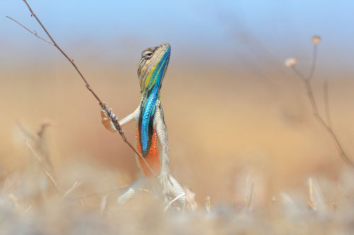
Warrior of the grassland - Anup Deodhar - The Comedy Wildlife.
Moonlight


-
 2op4au liked this · 7 years ago
 -
 mcm-curiosity liked this · 7 years ago
 -
 spacetimewithstuartgary reblogged this · 7 years ago
 -
 ricardelo reblogged this · 7 years ago
 -
 alxndrasplace reblogged this · 7 years ago
 -
 reporting-from-the-nerd-cave reblogged this · 7 years ago
 -
 salekinj reblogged this · 7 years ago
 -
salekinj liked this · 7 years ago
-
 votanh liked this · 7 years ago
 -
 realcleverscience reblogged this · 7 years ago
 -
 yojfull liked this · 7 years ago
 -
 magkevin86 liked this · 7 years ago
 -
 truthandvirtues liked this · 7 years ago
 -
 jaunit liked this · 7 years ago
 -
 traveler733 reblogged this · 7 years ago
 -
 spice-melange reblogged this · 7 years ago
 -
spice-melange liked this · 7 years ago
-
 smparticle2 reblogged this · 7 years ago
 -
 flyingmonkeynumber9 reblogged this · 7 years ago
 -
 hibegrfit liked this · 7 years ago
 -
 argentarachnids reblogged this · 7 years ago
 -
jackknighton liked this · 7 years ago
-
 ismael1106-blog liked this · 7 years ago
 -
 allgoodnamearetaken liked this · 7 years ago
 -
 gianthands liked this · 7 years ago
 -
 ekeshnan liked this · 7 years ago
 -
 nihilizards reblogged this · 7 years ago
 -
nihilizards liked this · 7 years ago
-
flyingmonkeynumber9 liked this · 7 years ago
-
 magiccircuspig liked this · 7 years ago
 -
 enlightened-logical-reliabl-blog liked this · 7 years ago
 -
 panda-of-the-trashh liked this · 7 years ago
 -
 joatgoog reblogged this · 7 years ago
 -
joatgoog liked this · 7 years ago
-
 sorrel-ly liked this · 7 years ago
 -
 stopthatitssilly reblogged this · 7 years ago
 -
 rahkshirock reblogged this · 7 years ago
 -
 redhousehead liked this · 7 years ago
 -
 andyt94-blog1 liked this · 7 years ago
 -
 wannabelehrer reblogged this · 7 years ago
 -
wannabelehrer liked this · 7 years ago
