It’s A Terrible Thing, I Think, In Life To Wait Until You’re Ready. I Have This Feeling Now That
It’s a terrible thing, I think, in life to wait until you’re ready. I have this feeling now that actually no one is ever ready to do anything. There is almost no such thing as ready. There is only now. And you may as well do it now.
Hugh Laurie (via liberatingreality)
More Posts from Smparticle2 and Others
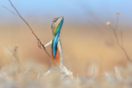
Warrior of the grassland - Anup Deodhar - The Comedy Wildlife.


GUYS https://twitter.com/AltNatParkSer/status/824054953404669953 http://www.scientistsmarchonwashington.com/ THE NATIONAL PARK SERVICE IS IN OPEN REBELLION


Rabies Viruses Reveal Wiring in Transparent Brains
Scientists under the leadership of the University of Bonn have harnessed rabies viruses for assessing the connectivity of nerve cell transplants. Coupled with a green fluorescent protein, the viruses show where replacement cells engrafted into mouse brains have connected to the host neural network.
The research is in Nature Communications. (full open access)

In slow motion, vortex rings can be truly stunning. This video shows two bubble rings underwater as they interact with one another. Upon approach, the two low-pressure vortex cores link up in what’s known as vortex reconnection. Note how the vortex rings split and reconnect in two places – not one. According to Helmholtz’s second theorem a vortex cannot end in a fluid–it must form a closed path (or end at a boundary); that’s why both sides come apart and together this way. After reconnection, waves ripple back and forth along the distorted vortex ring; these are known as Kelvin waves. Some of those perturbations bring two sides of the enlarged vortex ring too close to one another, causing a second vortex reconnection, which pinches off a smaller vortex ring. (Image source: A. Lawrence; submitted by Kam-Yung Soh)
Note: As with many viral images, locating a true source for this video is difficult. So far the closest to an original source I’ve found is the Instagram post linked above. If you know the original source, please let me know so that I can update the credit accordingly. Thanks!
NSF’s Science360 Photo of the Week
This is a close-up view of the beam created by a vortex laser.

Because the laser beam travels in a corkscrew pattern, encoding information into different vortex twists, it’s able to carry 10 times or more the amount of information than that of conventional lasers. The optics advancement could become a central component of next-generation computers designed to handle society’s growing demand for information sharing. Image credit: Natalia Litchinitser, University at Buffalo
Like this photo? Sign up for NSF’s Science360 News Service at news.science360.gov for a daily dose of STEM radio, news, videos and more cool images like this.

Women at work on a C-47 Douglas cargo transport, Douglas Aircraft Company, Long Beach, California, 1943.
via reddit

(Image caption: Brain showing hallmarks of Alzheimer’s disease (plaques in blue). Credit: ZEISS Microscopy)
New imaging technique measures toxicity of proteins associated with Alzheimer’s and Parkinson’s diseases
Researchers have developed a new imaging technique that makes it possible to study why proteins associated with Alzheimer’s and Parkinson’s diseases may go from harmless to toxic. The technique uses a technology called multi-dimensional super-resolution imaging that makes it possible to observe changes in the surfaces of individual protein molecules as they clump together. The tool may allow researchers to pinpoint how proteins misfold and eventually become toxic to nerve cells in the brain, which could aid in the development of treatments for these devastating diseases.
The researchers, from the University of Cambridge, have studied how a phenomenon called hydrophobicity (lack of affinity for water) in the proteins amyloid-beta and alpha synuclein – which are associated with Alzheimer’s and Parkinson’s respectively – changes as they stick together. It had been hypothesised that there was a link between the hydrophobicity and toxicity of these proteins, but this is the first time it has been possible to image hydrophobicity at such high resolution. Details are reported in the journal Nature Communications.
“These proteins start out in a relatively harmless form, but when they clump together, something important changes,” said Dr Steven Lee from Cambridge’s Department of Chemistry, the study’s senior author. “But using conventional imaging techniques, it hasn’t been possible to see what’s going on at the molecular level.”
In neurodegenerative diseases such as Alzheimer’s and Parkinson’s, naturally-occurring proteins fold into the wrong shape and clump together into filament-like structures known as amyloid fibrils and smaller, highly toxic clusters known as oligomers which are thought to damage or kill neurons, however the exact mechanism remains unknown.
For the past two decades, researchers have been attempting to develop treatments which stop the proliferation of these clusters in the brain, but before any such treatment can be developed, there first needs to be a precise understanding of how oligomers form and why.
“There’s something special about oligomers, and we want to know what it is,” said Lee. “We’ve developed new tools that will help us answer these questions.”
When using conventional microscopy techniques, physics makes it impossible to zoom in past a certain point. Essentially, there is an innate blurriness to light, so anything below a certain size will appear as a blurry blob when viewed through an optical microscope, simply because light waves spread when they are focused on such a tiny spot. Amyloid fibrils and oligomers are smaller than this limit so it’s very difficult to directly visualise what is going on.
However, new super-resolution techniques, which are 10 to 20 times better than optical microscopes, have allowed researchers to get around these limitations and view biological and chemical processes at the nanoscale.
Lee and his colleagues have taken super-resolution techniques one step further, and are now able to not only determine the location of a molecule, but also the environmental properties of single molecules simultaneously.
Using their technique, known as sPAINT (spectrally-resolved points accumulation for imaging in nanoscale topography), the researchers used a dye molecule to map the hydrophobicity of amyloid fibrils and oligomers implicated in neurodegenerative diseases. The sPAINT technique is easy to implement, only requiring the addition of a single transmission diffraction gradient onto a super-resolution microscope. According to the researchers, the ability to map hydrophobicity at the nanoscale could be used to understand other biological processes in future.
Spinal Stimulators Repurposed to Restore Touch in Lost Limb
Imagine tying your shoes or taking a sip of coffee or cracking an egg but without any feeling in your hand. That’s life for users of even the most advanced prosthetic arms.
Although it’s possible to simulate touch by stimulating the remaining nerves in the stump after an amputation, such a surgery is highly complex and individualized. But according to a new study from the University of Pittsburgh’s Rehab Neural Engineering Labs, spinal cord stimulators commonly used to relieve chronic pain could provide a straightforward and universal method for adding sensory feedback to a prosthetic arm.
For this study, published in eLife, four amputees received spinal stimulators, which, when turned on, create the illusion of sensations in the missing arm.

“What’s unique about this work is that we’re using devices that are already implanted in 50,000 people a year for pain — physicians in every major medical center across the country know how to do these surgical procedures — and we get similar results to highly specialized devices and procedures,” said study senior author Lee Fisher, Ph.D., assistant professor of physical medicine and rehabilitation, University of Pittsburgh School of Medicine.
The strings of implanted spinal electrodes, which Fisher describes as about the size and shape of “fat spaghetti noodles,” run along the spinal cord, where they sit slightly to one side, atop the same nerve roots that would normally transmit sensations from the arm. Since it’s a spinal cord implant, even a person with a shoulder-level amputation can use this device
Fisher’s team sent electrical pulses through different spots in the implanted electrodes, one at a time, while participants used a tablet to report what they were feeling and where.
All the participants experienced sensations somewhere on their missing arm or hand, and they indicated the extent of the area affected by drawing on a blank human form. Three participants reported feelings localized to a single finger or part of the palm.
“I was pretty surprised at how small the area of these sensations were that people were reporting,” Fisher said. “That’s important because we want to generate sensations only where the prosthetic limb is making contact with objects.”
When asked to describe not just where but how the stimulation felt, all four participants reported feeling natural sensations, such as touch and pressure, though these feelings often were mixed with decidedly artificial sensations, such as tingling, buzzing or prickling.
Although some degree of electrode migration is inevitable in the first few days after the leads are implanted, Fisher’s team found that the electrodes, and the sensations they generated, mostly stayed put across the month-long duration of the experiment. That’s important for the ultimate goal of creating a prosthetic arm that provides sensory feedback to the user.
“Stability of these devices is really critical,” Fisher said. “If the electrodes are moving around, that’s going to change what a person feels when we stimulate.”
The next big challenges are to design spinal stimulators that can be fully implanted rather than connecting to a stimulator outside the body and to demonstrate that the sensory feedback can help to improve the control of a prosthetic hand during functional tasks like tying shoes or holding an egg without accidentally crushing it. Shrinking the size of the contacts — the parts of the electrode where current comes out — is another priority. That might allow users to experience even more localized sensations.
“Our goal here wasn’t to develop the final device that someone would use permanently,” Fisher said. “Mostly we wanted to demonstrate the possibility that something like this could work.”
-
 femmeviva liked this · 1 year ago
 -
 alva-alice liked this · 1 year ago
 -
 potmirekom liked this · 1 year ago
 -
 lifesagame-playitwell reblogged this · 2 years ago
 -
 haileysun liked this · 3 years ago
 -
 daretothinkforyourself reblogged this · 3 years ago
 -
 karielyse1 reblogged this · 4 years ago
 -
 surz liked this · 4 years ago
 -
 satan-mistress liked this · 4 years ago
 -
 arxbqueen liked this · 5 years ago
 -
 non-existent3 reblogged this · 5 years ago
 -
 sinestezijaa reblogged this · 5 years ago
 -
sinestezijaa liked this · 5 years ago
-
 moon-kin reblogged this · 5 years ago
 -
daretothinkforyourself reblogged this · 5 years ago
-
 xlokisicequeenx reblogged this · 5 years ago
 -
xlokisicequeenx liked this · 5 years ago
-
 nowgostandinthecorner reblogged this · 5 years ago
 -
 elle-veee reblogged this · 6 years ago
 -
 nellvee reblogged this · 6 years ago
 -
 dynamicdumbledore liked this · 6 years ago
 -
 emilykg14 liked this · 6 years ago
 -
 electricsins reblogged this · 6 years ago
 -
 traced-in-blood reblogged this · 6 years ago
 -
 wannabezombiex liked this · 6 years ago
 -
 tanshiii liked this · 6 years ago
 -
 sad-but-rad18 liked this · 6 years ago
 -
 sippinlsd liked this · 6 years ago
 -
 zeit-geisty liked this · 6 years ago
 -
 maqdavlena reblogged this · 6 years ago
 -
 got-you-where-i-want-you reblogged this · 6 years ago
 -
 beauethereal reblogged this · 6 years ago
 -
 euphoricgemini reblogged this · 6 years ago
 -
 ecirtaebeinnab reblogged this · 6 years ago
 -
 fuckoffme247 reblogged this · 6 years ago
 -
fuckoffme247 liked this · 6 years ago
-
 40ouncesofgarbage reblogged this · 6 years ago
