CRISPR In Animals- Simplified. CRISPR Is A Breakthrough That Makes Genetic Editing Easier Than Ever Before.
CRISPR in Animals- Simplified. CRISPR is a breakthrough that makes genetic editing easier than ever before. How does it work, and what can it do? You can learn the basics in under 5 minutes from this animation.
More Posts from Contradictiontonature and Others
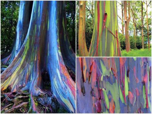
These are rainbow eucalyptus trees (Eucalyptus deglupta) and hail from the Philippine Islands.
The trees get their name from the striking colours observed on their trunks and limbs. Although it may look like someone took a paintbrush to them, these colours are entirely natural. Unlike most trees, the rainbow eucalyptus does not have a thick, cork-like layer of bark on its trunk. The bark is smooth and as it grows it ‘exfoliates’ layers of spent tissue. This exfoliation technique occurs at different stages and in different zones of the tree.
Keep reading

We might think we know the human body pretty well by now, but scientists are still discovering incredible individuals who are defying all odds by living out their lives with crucial parts missing, added, or tweaked in the most extraordinary ways.
From those with almost superhuman abilities, to others living without the organs we hold most dear, here are five of the most remarkable humans known to medicine.
Read more…


history meme (french edition) → 7 inventions/achievements (2/7) the first vaccine for rabies by Louis Pasteur & Émile Roux
“Pasteur had, in the early 1880s, a vaccine for rabies, but he was a chemist and not a licensed physician, and potentially liable if he injured or killed a human being. In early july 1885, Joseph Meister, a nine year-old-boy, had been badly mauled and bitten by a rabid dog (…). Pasteur injected young Meister with his rabies vaccine: the boy did not develop rabies and recovered fully from his injuries. Pasteur became a hero, and the Parisian Institue which came to be named in his honour, and of which he was the first director, became the global prototype bacteriological and immunological research institute. By demonstrating beyond doubt that many diseases were transmitted by bacteria and could be prevented from becoming active by pasteurization techniques, Pasteur indeed changed the course of history.” – G. L. Geison, The Private Science of Louis Pasteur.

Today, scientists working with telescopes at the European Southern Observatory and NASA announced a remarkable new discovery: An entire system of Earth-sized planets. If that’s not enough, the team asserts that the density measurements of the planets indicates that the six innermost are Earth-like rocky worlds.
And that’s just the beginning.
Three of the planets lie in the star’s habitable zone. If you aren’t familiar with the term, the habitable zone (also known as the “goldilocks zone”) is the region surrounding a star in which liquid water could theoretically exist. This means that all three of these alien worlds may have entire oceans of water, dramatically increasing the possibility of life. The other planets are less likely to host oceans of water, but the team states that liquid water is still a possibility on each of these worlds.
Summing the work, lead author Michaël Gillon notes that this solar system has the largest number of Earth-sized planets yet found and the largest number of worlds that could support liquid water: “This is an amazing planetary system — not only because we have found so many planets, but because they are all surprisingly similar in size to the Earth!”
Co-author Amaury Triaud notes that the star in this system is an “ultracool dwarf,” and he clarifies what this means in relation to the planets: “The energy output from dwarf stars like TRAPPIST-1 is much weaker than that of our Sun. Planets would need to be in far closer orbits than we see in the Solar System if there is to be surface water. Fortunately, it seems that this kind of compact configuration is just what we see around TRAPPIST-1.”
Continue Reading.
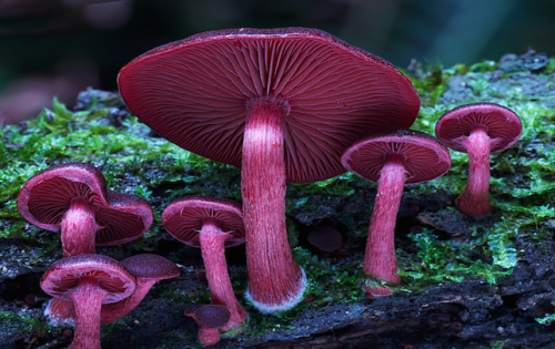
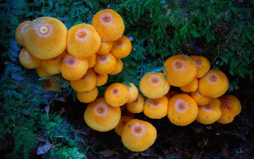
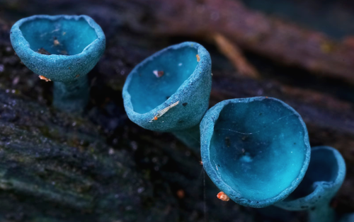
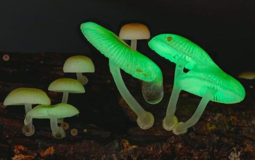

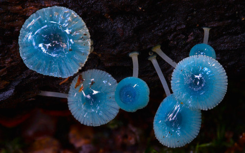
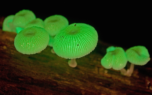
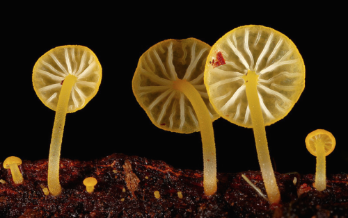
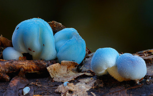
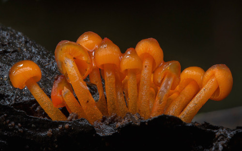
a mushroom rainbow to put the fun in fungi. cause they don’t need psilocybin to be magic. and though some mushrooms are coloured as a toxicity warning to predators, many others are brightly coloured to instead attract potential spore dispersers. see this for more on the bioluminescent mushrooms seen here. (photos)

For those poorly informed (educated) who insist that vaccines are just the same as catching the illness…. This is just one example of why that is not true.
Breakthrough for vaccine research: Mucosa forms special immunological memory
If a vaccine is to protect the intestines and other mucous membranes in the body, it also needs to be given through the mucosa, for example as a nasal spray or a liquid that is drunk. The mucosa forms a unique immunological antibody memory that does not occur if the vaccine is given by injection. This has been shown by a new study from Sahlgrenska Academy published in Nature Communications.
Immunological memory is the secret to human protection against various diseases and the success of vaccines. It allows our immune system to quickly recognize and neutralize threats. “The largest part of the immune system is in our mucosa. Even so, we understand less about how immunological memory protects us there than we do about protection in the rest of the body. Some have even suggested that a typical immune memory function does not exist in the mucosa,” says Mats Bemark, associate professor of immunology at Sahlgrenska Academy, University of Gothenburg.
After extensive work, the research team at Sahlgrenska Academy can now show that this assumption is completely wrong.
Mats Bemark et al. Limited clonal relatedness between gut IgA plasma cells and memory B cells after oral immunization, Nature Communications (2016). DOI: 10.1038/ncomms12698

A New Way to Cross the Blood–Brain Barrier - A Mental Unblock
The brain presents a unique challenge for medical treatment: it is locked away behind an impenetrable layer of tightly packed cells. Although the blood-brain barrier prevents harmful chemicals and bacteria from reaching our control center, it also blocks roughly 95 percent of medicine delivered orally or intravenously. As a result, doctors who treat patients with neurodegenerative diseases, such as Parkinson’s, often have to inject drugs directly into the brain, an invasive approach that requires drilling into the skull.
Some scientists have had minor successes getting intravenous drugs past the barrier with the help of ultrasound or in the form of nanoparticles, but those methods can target only small areas. Now neuroscientist Viviana Gradinaru and her colleagues at the California Institute of Technology show that a harmless virus can pass through the barricade and deliver treatment throughout the brain.
Gradinaru’s team turned to viruses because the infective agents are small and adept at entering cells and hijacking the DNA within. They also have protein shells that can hold beneficial deliveries, such as drugs or genetic therapies. To find a suitable virus to enter the brain, the researchers engineered a strain of an adeno-associated virus into millions of variants with slightly different shell structures. They then injected these variants into a mouse and, after a week, recovered the strains that made it into the brain. A virus named AAV-PHP.B most reliably crossed the barrier.
Next the team tested to see if AAV-PHP.B could work as a potential vector for gene therapy, a technique that treats diseases by introducing new genes into cells or by replacing or inactivating genes already there. The scientists injected the virus into the bloodstream of a mouse. In this case, the virus was carrying genes that encoded green fluorescent proteins. So if the virus made it to the brain and the new DNA was incorporated in neurons, the success rate could be tracked via a green glow on dissection. Indeed, the researchers observed that the virus infiltrated most brain cells and that the glowing effects lasted as long as one year. The results were recently published in Nature Biotechnology.
In the future, this approach could be used to treat a range of neurological diseases. “The ability to deliver genes to the brain without invasive methods will be extremely useful as a research tool. It has tremendous potential in the clinic as well,” says Anthony Zador, a neuroscientist who studies brain wiring at Cold Spring Harbor Laboratory. Gradinaru also thinks the method is a good candidate for targeting areas other than the brain, such as the peripheral nervous system. The sheer number of peripheral nerves has made pain treatment for neuropathy difficult, and a virus could infiltrate them all.
Image Credit: Thomas Fuchs
Source: Scientific American (By Monique Brouillette)

What Happens in the Brain During Unconsciousness?
Researchers are shining a light on the darkness of the unconscious brain. Three new studies add to the body of knowledge.
When patients undergo major surgery, they’re often put under anesthesia to allow the brain to be in an unconscious state.
But what’s happening in the brain during that time?
Three Michigan Medicine researchers are authors on three new articles from the Center for Consciousness Science exploring this question — specifically how brain networks fragment in association with a variety of unconsciousness states.
“These studies come from a long-standing hypothesis my colleagues and I have had regarding the essential characteristic of why we are conscious and how we become unconscious, based on patterns of information transfer in the brain,” says George A. Mashour, M.D., Ph.D., professor of anesthesiology, director of the Center for Consciousness Science and associate dean for clinical and translational research at the University of Michigan Medical School.
In the studies, the team not only explores how the brain networks fragment, but also how better to measure what is happening.
“We’ve been working for a decade to understand in a more refined way how the spatial and temporal aspects of brain function break down during unconsciousness, how we can measure that breakdown and the implications for information processing,” says UnCheol Lee, Ph.D., physicist, assistant professor of anesthesiology and associate director of the Center for Consciousness Science.
Examining different aspects of unconsciousness
The basis for the three studies, as well as other work from the Center for Consciousness Science, comes from a theory Mashour produced during his residency.
“I published a theoretical article when I was a resident in anesthesiology suggesting that anesthesia doesn’t work by turning the brain off, per se, but rather by isolating processes in certain areas of the brain,” Mashour says. “Instead of seeing a highly connected brain network, anesthesia results in an array of islands with isolated cognition and processing. We have taken this thought, as well as the work of others, and built upon it with our research.”
In the study in the Journal of Neuroscience, the team analyzed different areas of the brain during sedation, surgical anesthesia and a vegetative state.
“It’s often suggested that different areas of the brain that typically talk to one another get out of sync during unconsciousness,” says Anthony Hudetz, Ph.D., professor of anesthesiology, scientific director of the Center for Consciousness Science and senior author on the study. “We showed in the early stages of sedation, the information processing timeline gets much longer and local areas of the brain become more tightly connected within themselves. That tightening might lead to the inability to connect with distant areas.”
In the Frontiers in Human Neuroscience study, the team delved into how the brain integrates information and how it can be measured in the real world.
“We took a very complex computational task of measuring information integration in the brain and broke it down into a more manageable task,” says Lee, senior author on the study. “We demonstrated that as the brain gets more modular and has more local conversations, the measure of information integration starts to decrease. Essentially, we looked at how the brain network fragmentation was taking place and how to measure that fragmentation, which gives us the sense of why we lose consciousness.”
Finally, the latest article, in Trends in Neurosciences, aimed to take the team’s previous studies and other work on the subject of unconsciousness and put together a fuller picture.
“We examined unconsciousness across three different conditions: physiological, pharmacological and pathological,” says Mashour, lead author on the study. “We found that during unconsciousness, disrupted connectivity in the brain and greater modularity are creating an environment that is inhospitable to the kind of efficient information transfer that is required for consciousness.”
How these studies can help patients
The team members at the Center for Consciousness Science note that all of this work may help patients in the future.
“We’re looking for a better way to quantify the depth of anesthesia in the operating room and to assess consciousness in someone who has had a stroke or brain damage,” Hudetz says. “For example, we may assume that a patient is fully unconscious based on behavior, but in some cases consciousness can persist despite unresponsiveness.”
The team hopes this and future research could lead to therapeutic strategies for patients.
“We want to understand the communication breakdown that occurs in the brain during unconsciousness so we can precisely target or monitor these circuits to achieve safer anesthesia and restore these circuits to improve outcomes of coma,” Mashour says.

The Portuguese man o’ war delivers a powerful sting to its prey—and sometimes to people—through venom-filled structures on its tentacles. It is not a jellyfish, but rather a colony of different types of zooids (small animals). Jean Louis Coutant engraved the plate for this illustration.

Memory Competition
Most of the brain contains cells that no longer divide and renew. However, the dentate gyrus, nestled within the memory-forming centre of the brain (the hippocampus) is one of the few sites where new cells continue to form throughout life. As a person ages, there is an ever-increasing struggle for these new dentate gyrus neurons (coloured pink) to integrate with existing older neurons (green) because the latter already has well-established connections. This may be why learning and memorisation becomes more difficult as a person gets older. Scientists have now found that by temporarily reducing the number of dendritic spines – branches of neurons that form connections with other neurons – in the mature cells, the new cells have a better chance of functionally integrating. Indeed, in live mice, briefly eliminating dendritic spines boosted the number of integrated new neurons, which rejuvenated the hippocampus and improved the animals’ memory precision.
Written by Ruth Williams
Image courtesy of Kathleen McAvoy
Center for Regenerative Medicine, Massachusetts General Hospital, Boston, MA, USA
Copyright held by original authors
Research published in Neuron, September 2016
You can also follow BPoD on Twitter and Facebook
-
 toodlumx liked this · 3 years ago
 -
 vote-one-climb liked this · 4 years ago
 -
 soulrama liked this · 4 years ago
 -
 falconpunchyourmom reblogged this · 4 years ago
 -
 foggystudninja liked this · 4 years ago
 -
 day-knight reblogged this · 4 years ago
 -
day-knight liked this · 4 years ago
-
 robodoc250 liked this · 5 years ago
 -
 aleibah liked this · 5 years ago
 -
 nerdaces reblogged this · 5 years ago
 -
 ayumunoya liked this · 5 years ago
 -
 dreamyeyes26 liked this · 5 years ago
 -
 imperialcrystalgirl liked this · 5 years ago
 -
 angelicdomi liked this · 5 years ago
 -
 vixeyfoxwishdouglas reblogged this · 5 years ago
 -
vixeyfoxwishdouglas reblogged this · 5 years ago
-
 herbygrad reblogged this · 5 years ago
 -
herbygrad liked this · 5 years ago
-
 caterpillarfuture reblogged this · 5 years ago
 -
 handsforpuns liked this · 5 years ago
 -
 50ci3ty5uck5 reblogged this · 6 years ago
 -
 dontaskdonttelldontworry liked this · 6 years ago
 -
 harpylegs liked this · 6 years ago
 -
 stetskiwi liked this · 6 years ago
 -
 slushpussy reblogged this · 6 years ago
 -
 biology-physics-chemistry-math reblogged this · 6 years ago
 -
 quartzyblr reblogged this · 6 years ago
 -
quartzyblr liked this · 6 years ago
-
 sparechangeofheart liked this · 6 years ago
 -
 stvdyhoe liked this · 6 years ago
 -
 rottedwing liked this · 6 years ago
 -
 ashlienotashley liked this · 6 years ago
 -
 ladyluck25 liked this · 6 years ago
 -
 silencio0ldman liked this · 6 years ago
 -
 crispr-the-human-creation-kit reblogged this · 6 years ago
 -
 siamesefever liked this · 6 years ago
A pharmacist and a little science sideblog. "Knowledge belongs to humanity, and is the torch which illuminates the world." - Louis Pasteur
215 posts