contradictiontonature
A pharmacist and a little science sideblog. "Knowledge belongs to humanity, and is the torch which illuminates the world." - Louis Pasteur
215 posts
Latest Posts by contradictiontonature

For #WorldBeeDay, here’s a look at the chemistry behind the honey some bees produce: https://ift.tt/2GV5qtq https://ift.tt/2LJpsIe
Thyroid function tests

Standard Tests
TSH levels
Free T4 (fT4) levels
Measurements of total T4 + T3 used to be common however detects both bound and free T3 + T4
Elevated total T4 may occur in healthy individuals if there is an increase in binding protein concentrations
Reliable tests now exist for free T4 + T3
T3 = 3.9-6.7 pmol/L
T4 = 12-22 pmol/L
Thyroid-stimulating hormone
Produced by the pituitary gland, not the thyroid, however:
TSH levels are controlled by negative feedback – can be indication of thyroid function (changes in T3+T4 will result in changes in TSH to try compesate)
TSH levels greatly elevated in hypothyroidism – >10 fold increase over reference values
More sensitive marker than decreased fT4 - increased TSH occurs before fT4 decreases
TSH levels greatly supressed in hyperthyroidism
Low concentrations can also occur in non-thyroidal illness
TSH measurement is the first-line test of thyroid function.
Free T4 + T3 Measurements
Desirable as free hormone is clinically relevant
Total levels can change under conditions that alter thyroxine-binding globulin (TBG) levels e.g. pregnancy
Large changes in TBG may still affect fT4 + fT3 levels
fT3 levels often normal in hypothyroidism
fT3 levels usually raised more than fT4 levels in hyperthyroidism
Unless complicated by an illness effecting conversion of T4 to T3
Therefore: – fT4 levels are a better indication of hypothyroidism
fT3 levels are a better indication of hyperthyroidism


Hepatitis B in a Laboratory!! #serology #infection #liver #hepatitis #medicine #medstudent #medstudynotes #virus #medschool #microbiology #pathology https://www.instagram.com/p/BrIYa-LheyH/?utm_source=ig_tumblr_share&igshid=lqm0j92yobw0
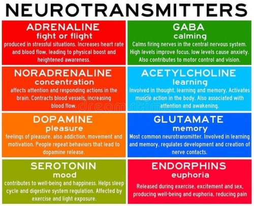
Neurotransmitters are chemicals that help in transmitting signals across a synapse. Different neurotransmitters are associated with different functions. Knowledge about these helps us to treat various neurological conditions by either stimulating or inhibiting these production. #neurology #neuroscience #psychiatry #medicine #medstudynotes #medschool #mbbs #unimed #brain #nervoussystem #physiology #medblog #medblr #medstudent https://www.instagram.com/p/BrM4ocsBqJe/?utm_source=ig_tumblr_share&igshid=12tojib83c32d

January is #NationalBloodDonorMonth 💉 Which different blood types are compatible with one another? This graphic takes a look: https://ift.tt/35wB7S9 https://ift.tt/2ugvtXs

How Do Antidepressants Work? (Video)
Your brain is a network of billions of neurones, all somehow connected to each other. At this very second, millions of impulses are being transmitted through these connections carrying information about what you can see and hear, as well as your emotional state. It’s an incredibly complex system but sometimes things go wrong. Despite extensive research, we are still not certain on the biology that underlies mental illnesses- including depression. However, we have come pretty far in developing effective treatments.
Thorny life of new-born neurons
Even in adult brains, new neurons are generated throughout a lifetime. In a publication in the scientific journal PNAS, a research group led by Goethe University describes plastic changes of adult-born neurons in the hippocampus, a critical region for learning: frequent nerve signals enlarge the spines on neuronal dendrites, which in turn enables contact with the existing neural network.

Practise makes perfect, and constant repetition promotes the ability to remember. Researchers have been aware for some time that repeated electrical stimulation strengthens neuron connections (synapses) in the brain. It is similar to the way a frequently used trail gradually widens into a path. Conversely, if rarely used, synapses can also be removed – for example, when the vocabulary of a foreign language is forgotten after leaving school because it is no longer practised. Researchers designate the ability to change interconnections permanently and as needed as the plasticity of the brain.
Plasticity is especially important in the hippocampus, a primary region associated with long-term memory, in which new neurons are formed throughout life. The research groups led by Dr Stephan Schwarzacher (Goethe University), Professor Peter Jedlicka (Goethe University and Justus Liebig University in Gießen) and Dr Hermann Cuntz (FIAS, Frankfurt) therefore studied the long-term plasticity of synapses in new-born hippocampal granule cells. Synaptic interconnections between neurons are predominantly anchored on small thorny protrusions on the dendrites called spines. The dendrites of most neurons are covered with these spines, similar to the thorns on a rose stem.
In their recently published work, the scientists were able to demonstrate for the first time that synaptic plasticity in new-born neurons is connected to long-term structural changes in the dendritic spines: repeated electrical stimulation strengthens the synapses by enlarging their spines. A particularly surprising observation was that the overall size and number of spines did not change: when the stimulation strengthened a group of synapses, and their dendritic spines enlarged, a different group of synapses that were not being stimulated simultaneously became weaker and their dendritic spines shrank.
“This observation was only technically possible because our students Tassilo Jungenitz and Marcel Beining succeeded for the first time in examining plastic changes in stimulated and non-stimulated dendritic spines within individual new-born cells using 2-photon microscopy and viral labelling,” says Stephan Schwarzacher from the Institute for Anatomy at the University Hospital Frankfurt. Peter Jedlicka adds: “The enlargement of stimulated synapses and the shrinking of non-stimulated synapses was at equilibrium. Our computer models predict that this is important for maintaining neuron activity and ensuring their survival.”
The scientists now want to study the impenetrable, spiny forest of new-born neuron dendrites in detail. They hope to better understand how the equilibrated changes in dendritic spines and their synapses contribute the efficient storing of information and consequently to learning processes in the hippocampus.
Complement Pathways

Components of complement pathways of the immune system.
Classical Pathway: binds to the pathogen surface
C1 binds to phosphocholine on bacteria, which activates C1r to cleave C1s.
Activated C1s cleaves C4 to C4a and C4b.
C4b binds to the microbial surface and also binds C2.
C2 is cleaved to C2a and C2b by C1s, forming the C4bC2a complex.
The C4bC2a complex cleaves C3 to C3a and C3b.
C3b binds to the surface and causes opsonization.
MB-Lectin Pathway: uses mannin-binding lectin to bind to mannose-containing carbohydrates on the pathogen surface
Mannin-binding lectin (MBL) binds to the pathogen surface and activates MASP-2.
MASP-2 cleaves C4 to C4a and C4b.
C4b binds to the microbial surface and also binds C2.
C2 is cleaved to C2a and C2b by MASP-2, forming the C4bC2a complex.
The C4bC2a complex cleaves C3 to C3a and C3b.
C3b binds to the surface and causes opsonization.
Alternative Pathway: binds to the pathogen surface with spontaneously activated complement, amplifies C3b
C3b deposited by the C3 convertase binds to factor B.
Factor B is cleaved by factor D into Ba and Bb, forming the C3bBb complex.
The C3bBb complex cleaves C3 into C3a and C3b.
C3 spontaneously hydrolyzes to C3(H2O).
C3(H2O) binds to factor B, and factor D cleaves factor B.
Upon factor B cleavage, the C3(H2O)Bb complex is formed.
The C3(H2O)Bb complex cleaves C3 into C3a and C3b.
Factor B binds to C3b on the surface and is cleaved to Bb.


Liver Circulation - Flashcard

The liver is supplied with blood by the hepatic artery and the hepatic portal vein
branches of the hepatic artery and the hepatic portal vein distribute blood to the periphery of the liver lobules.
Blood passes along sinusoids, which are lined by hepatocytes, which perform numerous metabolic and synthetic functions.
The processed blood passes into branches of the hepatic vein in the centre of each lobule, and eventually drains into the hepatic vein.
The biliary system is independent of the vascular system and bile moves in the opposite direction to the blood.
Initially it is collected in bile ductules which are surrounded by collagenous tissue, which forms part of the collagenous trabecular septum.
The bile is collected by increasingly large trabecular ducts, which fuse to form intrahepatic ducts which finally drain into the main hepatic ducts.


Summary of Antibiotics.. #pharmacologyreview #pharmacology #pharmacologist #physiology #pathology #usmle #usmlestep1 #usmlestep2 #doctor #doctordconline #nhs #nurse #nursing #hospital #patient #mbbs #md #medicine @doctordconline


How are elements created in space, stars, and in laboratories? The latest edition of #PeriodicGraphics in C&EN takes a look! http://bit.ly/2UXWoPD http://bit.ly/2YbuBNE

Antibiotic resistance has been called one of the biggest public health threats of our time. There is a pressing need for new and novel antibiotics to combat the rise in antibiotic-resistant bacteria worldwide.
Researchers from Florida International University’s Herbert Wertheim College of Medicine are part of an international team that has discovered a new broad-spectrum antibiotic that contains arsenic. The study, published in Nature’s Communication Biology, is a collaboration between Barry P. Rosen, Masafumi Yoshinaga, Venkadesh Sarkarai Nadar and others from the Department of Cellular Biology and Pharmacology, and Satoru Ishikawa and Masato Kuramata from the Institute for Agro-Environmental Sciences, NARO in Japan.
“The antibiotic, arsinothricin or AST, is a natural product made by soil bacteria and is effective against many types of bacteria, which is what broad-spectrum means,” said Rosen, co-senior author of the study published in the Nature journal, Communications Biology. “Arsinothricin is the first and only known natural arsenic-containing antibiotic, and we have great hopes for it.”
Although it contains arsenic, researchers say they tested AST toxicity on human blood cells and reported that “it doesn’t kill human cells in tissue culture.”
Continue Reading.
Gertrude B. Elion: Women who changed science
With the drugs that she created, Gertrude Elion fulfilled her life’s mission: to alleviate human suffering. Beyond the individual drugs she discovered, she pioneered a new, more scientific approach to drug development that forever altered – and accelerated – medical research.
Discover her story at
https://www.nobelprize.org/womenwhochangedscience/stories


Breaking News:
The Nobel Prize in Physics for 2018 has been awarded to Arthur Ashkin, Gerard Mourou and Donna Strickland “for groundbreaking inventions in the field of laser physics”.
Donna Strickland is the first woman to win the Nobel Prize in Physics in 55 years.
Nobel Laureate Arthur Ashkin has been awarded the #NobelPrize in Physics “for the optical tweezers and their application to biological systems.”
Nobel Laureates Gérard Mourou and Donna Strickland have been awarded the #NobelPrize in Physics “for their method of generating high-intensity, ultra-short optical pulses.”
Article here with more information about their work:
Arthur Ashkin, Gérard Mourou and Donna Strickland win Nobel physics prize

Social Stress Leads To Changes In Gut Bacteria
Exposure to psychological stress in the form of social conflict alters gut bacteria in Syrian hamsters, according to a new study by Georgia State University.
It has long been said that humans have “gut feelings” about things, but how the gut might communicate those “feelings” to the brain was not known. It has been shown that gut microbiota, the complex community of microorganisms that live in the digestive tracts of humans and other animals, can send signals to the brain and vice versa.
In addition, recent data have indicated that stress can alter the gut microbiota. The most common stress experienced by humans and other animals is social stress, and this stress can trigger or worsen mental illness in humans. Researchers at Georgia State have examined whether mild social stress alters the gut microbiota in Syrian hamsters, and if so, whether this response is different in animals that “win” compared to those that “lose” in conflict situations.
Hamsters are ideal to study social stress because they rapidly form dominance hierarchies when paired with other animals. In this study, pairs of adult males were placed together and they quickly began to compete, resulting in dominant (winner) and subordinate (loser) animals that maintained this status throughout the experiment. Their gut microbes were sampled before and after the first encounter as well as after nine interactions. Sampling was also done in a control group of hamsters that were never paired and thus had no social stress. The researchers’ findings are published in the journal Behavioural Brain Research.
“We found that even a single exposure to social stress causes a change in the gut microbiota, similar to what is seen following other, much more severe physical stressors, and this change gets bigger following repeated exposures,” said Dr. Kim Huhman, Distinguished University Professor of Neuroscience at Georgia State. “Because ‘losers’ show much more stress hormone release than do ‘winners,’ we initially hypothesized that the microbial changes would be more pronounced in animals that lost than in animals that won.”
“Interestingly, we found that social stress, regardless of who won, led to similar overall changes in the microbiota, although the particular bacteria that were impacted were somewhat different in winners and losers. It might be that the impact of social stress was somewhat greater for the subordinate animals, but we can’t say that strongly.”
Another unique finding came from samples that were taken before the animals were ever paired, which were used to determine if any of the preexisting bacteria seemed to correlate with whether an animal turned out to be the winner or loser.
“It’s an intriguing finding that there were some bacteria that seemed to predict whether an animal would become a winner or a loser,” Huhman said.
“These findings suggest that bi-directional communication is occurring, with stress impacting the microbiota, and on the other hand, with some specific bacteria in turn impacting the response to stress,” said Dr. Benoit Chassaing, assistant professor in the Neuroscience Institute at Georgia State.
This is an exciting possibility that builds on evidence that gut microbiota can regulate social behavior and is being investigated by Huhman and Chassaing.

What Happens in the Brain During Unconsciousness?
Researchers are shining a light on the darkness of the unconscious brain. Three new studies add to the body of knowledge.
When patients undergo major surgery, they’re often put under anesthesia to allow the brain to be in an unconscious state.
But what’s happening in the brain during that time?
Three Michigan Medicine researchers are authors on three new articles from the Center for Consciousness Science exploring this question — specifically how brain networks fragment in association with a variety of unconsciousness states.
“These studies come from a long-standing hypothesis my colleagues and I have had regarding the essential characteristic of why we are conscious and how we become unconscious, based on patterns of information transfer in the brain,” says George A. Mashour, M.D., Ph.D., professor of anesthesiology, director of the Center for Consciousness Science and associate dean for clinical and translational research at the University of Michigan Medical School.
In the studies, the team not only explores how the brain networks fragment, but also how better to measure what is happening.
“We’ve been working for a decade to understand in a more refined way how the spatial and temporal aspects of brain function break down during unconsciousness, how we can measure that breakdown and the implications for information processing,” says UnCheol Lee, Ph.D., physicist, assistant professor of anesthesiology and associate director of the Center for Consciousness Science.
Examining different aspects of unconsciousness
The basis for the three studies, as well as other work from the Center for Consciousness Science, comes from a theory Mashour produced during his residency.
“I published a theoretical article when I was a resident in anesthesiology suggesting that anesthesia doesn’t work by turning the brain off, per se, but rather by isolating processes in certain areas of the brain,” Mashour says. “Instead of seeing a highly connected brain network, anesthesia results in an array of islands with isolated cognition and processing. We have taken this thought, as well as the work of others, and built upon it with our research.”
In the study in the Journal of Neuroscience, the team analyzed different areas of the brain during sedation, surgical anesthesia and a vegetative state.
“It’s often suggested that different areas of the brain that typically talk to one another get out of sync during unconsciousness,” says Anthony Hudetz, Ph.D., professor of anesthesiology, scientific director of the Center for Consciousness Science and senior author on the study. “We showed in the early stages of sedation, the information processing timeline gets much longer and local areas of the brain become more tightly connected within themselves. That tightening might lead to the inability to connect with distant areas.”
In the Frontiers in Human Neuroscience study, the team delved into how the brain integrates information and how it can be measured in the real world.
“We took a very complex computational task of measuring information integration in the brain and broke it down into a more manageable task,” says Lee, senior author on the study. “We demonstrated that as the brain gets more modular and has more local conversations, the measure of information integration starts to decrease. Essentially, we looked at how the brain network fragmentation was taking place and how to measure that fragmentation, which gives us the sense of why we lose consciousness.”
Finally, the latest article, in Trends in Neurosciences, aimed to take the team’s previous studies and other work on the subject of unconsciousness and put together a fuller picture.
“We examined unconsciousness across three different conditions: physiological, pharmacological and pathological,” says Mashour, lead author on the study. “We found that during unconsciousness, disrupted connectivity in the brain and greater modularity are creating an environment that is inhospitable to the kind of efficient information transfer that is required for consciousness.”
How these studies can help patients
The team members at the Center for Consciousness Science note that all of this work may help patients in the future.
“We’re looking for a better way to quantify the depth of anesthesia in the operating room and to assess consciousness in someone who has had a stroke or brain damage,” Hudetz says. “For example, we may assume that a patient is fully unconscious based on behavior, but in some cases consciousness can persist despite unresponsiveness.”
The team hopes this and future research could lead to therapeutic strategies for patients.
“We want to understand the communication breakdown that occurs in the brain during unconsciousness so we can precisely target or monitor these circuits to achieve safer anesthesia and restore these circuits to improve outcomes of coma,” Mashour says.

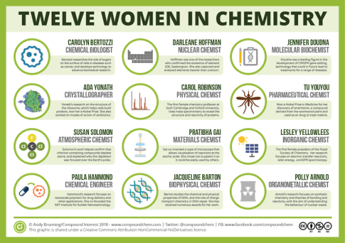
For International Women’s Day, here are 12 women from chemistry history: wp.me/p4aPLT-2ra and 12 from chemistry present: wp.me/p4aPLT-5w7

The first synthesis of aspirin was carried out #OTD in 1897. Here’s a look at how it compares to other painkillers: http://www.compoundchem.com/2014/09/25/painkillers/

Science Fact Friday: Tetrodotoxin, ft. a small gif because I’m avoiding my real obligations. Why does tetrodotoxin not affect its host? More studies need to be done but at least a few species possess mutated sodium ion channels. The tetrodotoxin can’t interact efficiently with the altered channels.
Another interesting tidbit: Animals with tetrodotoxin can lose their toxicity in captivity. It is suspected that the animals accumulate the toxic bacteria as a side-effect of their diet. After several years of captivity on a tetrodotoxin-bacteria-free diet, the bacterial colonies living in the animals die, residual toxin is cleared from the system, and the animal is safe to handle.
Estrogen Alters Memory Circuit Function in Women with Gene Variant
Fluctuations in estrogen can trigger atypical functioning in a key brain memory circuit in women with a common version of a gene, NIMH scientists have discovered. Brain scans revealed altered circuit activity linked to changes in the sex hormone in women with the gene variant while they performed a working memory task.

(Image caption: Both PET scans (left) and fMRI scans (right) showed the same atypical activation (yellow) in the brain’s memory hub, or hippocampus, in response to estrogen in women performing a working memory task – if they carried a uniquely human version of the BDNF gene. Activity in this area is typically suppressed during working memory. Picture shows PET and fMRI data superimposed over anatomical MRI image)
The findings may help to explain individual differences in menstrual cycle and reproductive-related mental disorders linked to fluctuations in the hormone. They may also shed light on mechanisms underlying sex-related differences in onset, severity, and course of mood and anxiety disorders and schizophrenia. The gene-by-hormone interaction’s effect on circuit function was found only with one of two versions of the gene that occurs in about a fourth of white women.
Drs. Karen Berman, Peter Schmidt, Shau-Ming Wei, and colleagues, of the NIMH Intramural Research Program, report on this first such demonstration in women April 18, 2017 in the journal Molecular Psychiatry.
Prior to the study, there was little evidence from research on the human brain that might account for individual differences in cognitive and behavioral effects of sex hormones. For example, why do some women develop postpartum depression and others do not – in response to the same hormone changes? Why do some women report that estrogen replacement improved their memory, whereas large studies of postmenopausal estrogen therapy show no overall improvement in memory performance?
Evidence from humans has also been lacking for the neural basis of stark sex differences in prevalence and course of mental disorders that are likely related to sex hormones. For example, why are there higher rates of mood disorders in females and higher rates of ADHD in males – or later onset of schizophrenia in females?
In seeking answers to these questions, the researchers focused on working memory, a well-researched brain function often disturbed in many of these disorders. It was known that working memory is mediated by a circuit from the brain’s executive hub, the prefrontal cortex, to its memory hub, the hippocampus. Notably, hippocampus activity is typically suppressed during working memory processing.
Following-up on a clue from experiments in mice, the NIMH team hypothesized that estrogen tweaks circuit function by interacting with a uniquely human version of the gene that codes for brain derived neurotrophic factor (BDNF), a pivotal chemical messenger operating in this circuit. To find out, the researchers experimentally manipulated estrogen levels in healthy women with one or the other version of the BDNF gene over a period of months. Researchers periodically scanned the women’s brain activity while they performed a working memory task to see any effects of the gene-hormone interaction on circuit function.
The researchers first scanned 39 women using PET (positron emission tomography) and later confirmed the results in 27 women using fMRI (functional magnetic resonance imaging). Both pegged atypical activity in the hippocampus to the interaction. Turning up the same findings using two types of neuroimaging strengthens the case for the accuracy of their observations, say the researchers. Such gene-hormone interactions affecting thinking and behavior are consistent with findings from animal studies and are suspect mechanisms conferring risk for mental illness, they add.

Today is the Autumn Equinox in the northern hemisphere! What’s behind the changing colours of autumn leaves? http://wp.me/p4aPLT-sn

Stephanie Kwolek was born #OTD in 1923. She’s most famous for inventing the bulletproof polymer Kevlar: wp.me/p4aPLT-3dv


Scientists are pretty sure that deep inside the moon, there’s water
While Earth’s surface cracks and spouts fire, the moon’s surface, for as long as we’ve known it, has been quiet.
The youngest sign of volcanic activity scientists have found on the moon’s surface is 18 million years old.
But the traces of that long-ago volcanic activity could help scientists crack an enduring mystery: How much water is on the moon?
A study published Monday in Nature Geoscience suggests it may be more than we thought. Read more (7/24/17)
follow @the-future-now

Rosalind Franklin was born #OTD in 1920. Her work was instrumental in the discovery of the structure of DNA: http://wp.me/s4aPLT-franklin