Collective Memory In Bacteria

Collective memory in bacteria
Individual bacterial cells have short memories. But groups of bacteria can develop a collective memory that can increase their tolerance to stress. This has been demonstrated experimentally for the first time in a study by Eawag and ETH Zurich scientists published in PNAS.
Roland Mathis, Martin Ackermann. Response of single bacterial cells to stress gives rise to complex history dependence at the population level. PNAS, March 7, 2016 DOI: 10.1073/pnas.1511509113
Experimental set-up with the bacterium Caulobacter crescentus in microfluidic chips: each chip comprises eight channels, with a bacterial population growing in each channel. The bacteria are attached to the glass surface by an adhesive stalk. When the bacterial cells divide, one of the two daughter cells remains in the channel, while the other is washed out. Using time-lapse microscopy, bacterial cell-division cycles and survival probabilities can thus be reconstructed. Credit: Stephanie Stutz
More Posts from Contradictiontonature and Others

(Image caption: The synapses of pyramid cells in the cerebral cortex form functional groups. Some of the related synapses are shown in green in the reconstruction. Credit: © MPI of Neurobiology / Scheuss)
Neurons form synapse clusters
The cerebral cortex resembles a vast switchboard. Countless lines carrying information about the environment, for example from the sensory organs, converge in the cerebral cortex. In order to direct the flow of data into meaningful pathways, the individual pyramidal cells of the cerebral cortex act like miniature switchboard operators. Each cell receives information from several thousand lines. If the signals make sense, the line is opened, and the information is relayed onward. Scientists at the Max Planck Institute of Neurobiology in Martinsried have now shown for the first time that contact points between specific neuron types are clustered in groups on the target neuron. It is probable that signals are coordinated with each other in this way to make them more “convincing”.
The cells of the cerebral cortex have a lot to do. They process various types of information depending on the area in which they are located. For example, signals from the retina arrive in the visual cortex, where, among other things, the motion of objects is detected. The pyramidal cells of the cerebral cortex receive information from other cells through thousands of contact points called synapses. Depending on where, how many and how often synapses are activated, the cell relays the signal onward – or not.
Information is passed on in the form of electrical signals. The neurobiologists were able to measure these signals at various contact points of the neuron. “The exciting thing is that the signals that a cell receives from, say, ten simultaneously active synapses can be greater than the sum of the signals from the ten individual synapses,” says Volker Scheuss, summarizing the basis of his recently published study. “However, until now it was unclear whether this phenomenon can be explained by a specific arrangement of synapses on pyramidal cells.”
By combining modern methods, the neurobiologists in Tobias Bonhoeffer’s Department have analysed the arrangement of synapses. They were able to selectively activate a specific type of pyramid cell in brain slices from mice using optogenetics. Thanks to simultaneous “calcium imaging”, they were then able to observe and record the activity of individual synapses under a two-photon microscope. In this way, they succeeded in showing for the first time how synapses are arranged with respect to each other.
The result of such synapse mapping analysed with a newly developed algorithm was clear: The synapses of pyramidal cells form clusters consisting of 4 to 14 synapses arranged within an area of less than 30 micrometres along the dendrite. “The existence of these clusters suggests that the synapses interact with each other to control the strength of the combined signal,” explains Onur Gökçe, author of the study. This is the first anatomical explanation for the disproportionate strength of clustered synapse signals in comparison to the individual signals – a finding known from activity measurements. The observation in layer 5 pyramidal cells was of particular interest, as the activity of these cells oscillates synchronously. “This rhythmic activity, which probably influences the processing of visual information, could synchronously activate synapse clusters, thus boosting the overall signal received,” says Scheuss.

At last, we’ve seen what might be the primary building blocks of memories lighting up in the brains of mice.
We have cells in our brains – and so do rodents – that keep track of our location and the distances we’ve travelled. These neurons are also known to fire in sequence when a rat is resting, as if the animal is mentally retracing its path – a process that probably helps memories form, says Rosa Cossart at the Institut de Neurobiologie de la Méditerranée in Marseille, France.
But without a way of mapping the activity of a large number of these individual neurons, the pattern that these replaying neurons form in the brain has been unclear. Researchers have suspected for decades that the cells might fire together in small groups, but nobody could really look at them, says Cossart.
To get a look, Cossart and her team added a fluorescent protein to the neurons of four mice. This protein fluoresces the most when calcium ions flood into a cell – a sign that a neuron is actively firing. The team used this fluorescence to map neuron activity much more widely than previous techniques, using implanted electrodes, have been able to do.
Observing the activity of more than 1000 neurons per mouse, the team watched what happened when mice walked on a treadmill or stood still.
As expected, when the mice were running, the neurons that trace how far the animal has travelled fired in a sequential pattern, keeping track.
These same cells also lit up while the mice were resting, but in a strange pattern. As they reflected on their memories, the neurons fired in the same sequence as they had when the animals were running, but much faster. And rather than firing in turn individually, they fired together in sequential blocks that corresponded to particular fragments of a mouse’s run.
“We’ve been able to image the individual building-blocks of memory,” Cossart says, each one reflecting a chunk of the original episode that the mouse experienced.
Continue Reading.

Today is the Autumn Equinox in the northern hemisphere! What’s behind the changing colours of autumn leaves? http://wp.me/p4aPLT-sn

Quote by #rosalindfranklin How do you make science a part of your life? What are you doing to fight for scientific literacy? More quotes and questions in my #ilovescience journal. #womeninscience #scientificliteracy

Viruses support photosynthesis in bacteria: An evolutionary advantage?
Viruses propagate by infecting a host cell and reproducing inside. This not only affects humans and animals, but bacteria as well. This type of virus is called bacteriophage. They carry so called auxiliary metabolic genes in their genome, which are responsible for producing certain proteins that give the virus an advantage. Researchers at the University of Kaiserslautern and the Ruhr University Bochum have analysed the structure of such a protein more closely. It appears to stimulate the photosynthesis of host bacteria. The study has now been published in the journal The Journal of Biological Chemistry.
Raphael Gasper, Julia Schwach, Jana Hartmann, Andrea Holtkamp, Jessica Wiethaus, Natascha Riedel, Eckhard Hofmann, Nicole Frankenberg-Dinkel. Auxiliary metabolic genes- Distinct Features of Cyanophage-encoded T-type Phycobiliprotein Lyase θCpeT. Journal of Biological Chemistry, 2017; jbc.M116.769703 DOI: 10.1074/jbc.M116.769703
The association between the virus protein and bacterial pigment is incredibly stable. Furthermore, the complex is highly fluorescent. Credit: AG Frankenberg-Dinkel

Memory Competition
Most of the brain contains cells that no longer divide and renew. However, the dentate gyrus, nestled within the memory-forming centre of the brain (the hippocampus) is one of the few sites where new cells continue to form throughout life. As a person ages, there is an ever-increasing struggle for these new dentate gyrus neurons (coloured pink) to integrate with existing older neurons (green) because the latter already has well-established connections. This may be why learning and memorisation becomes more difficult as a person gets older. Scientists have now found that by temporarily reducing the number of dendritic spines – branches of neurons that form connections with other neurons – in the mature cells, the new cells have a better chance of functionally integrating. Indeed, in live mice, briefly eliminating dendritic spines boosted the number of integrated new neurons, which rejuvenated the hippocampus and improved the animals’ memory precision.
Written by Ruth Williams
Image courtesy of Kathleen McAvoy
Center for Regenerative Medicine, Massachusetts General Hospital, Boston, MA, USA
Copyright held by original authors
Research published in Neuron, September 2016
You can also follow BPoD on Twitter and Facebook

Atomic-level motion may drive bacteria’s ability to evade immune system defenses
A study from Indiana University has found evidence that extremely small changes in how atoms move in bacterial proteins can play a big role in how these microorganisms function and evolve.
The research, recently published in the Proceedings of the National Academy of Sciences, is a major departure from prevailing views about the evolution of new functions in organisms, which regarded a protein’s shape, or “structure,” as the most important factor in controlling its activity.
“This study gives us a significant answer to the following question: How do different organisms evolve different functions with proteins whose structures all look essentially the same?” said David Giedroc, Lilly Chemistry Alumni Professor in the IU Bloomington College of Arts and Sciences’ Department of Chemistry, who is senior author on the study. “We’ve found evidence that atomic motions in proteins play a major role in impacting their function.”
Daiana A. Capdevila et al, Entropy redistribution controls allostery in a metalloregulatory protein, Proceedings of the National Academy of Sciences (2017). DOI: 10.1073/pnas.1620665114
The scientists conducted their experiments in Staphylococcus aureus, a common cause of skin, sinus and lung infections. Credit: Centers for Disease Control and Prevention
It is imperfection - not perfection - that is the end result of the program written into that formidably complex engine that is the human brain, and of the influences exerted upon us by the environment and whoever takes care of us during the long years of physical, psychological and intellectual development.
Rita Levi-Montalcini

Image Credit: Hammersmith Hospital in London
(via neuromorphogenesis)
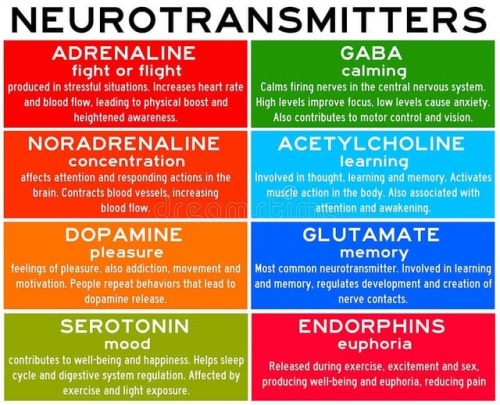
Neurotransmitters are chemicals that help in transmitting signals across a synapse. Different neurotransmitters are associated with different functions. Knowledge about these helps us to treat various neurological conditions by either stimulating or inhibiting these production. #neurology #neuroscience #psychiatry #medicine #medstudynotes #medschool #mbbs #unimed #brain #nervoussystem #physiology #medblog #medblr #medstudent https://www.instagram.com/p/BrM4ocsBqJe/?utm_source=ig_tumblr_share&igshid=12tojib83c32d


-
 xxglitterdalambxx liked this · 5 years ago
 -
 drjoebell reblogged this · 8 years ago
 -
 devastationwagon liked this · 8 years ago
 -
 contradictiontonature reblogged this · 8 years ago
 -
 zenosanalytic liked this · 8 years ago
 -
 manyblinkinglights liked this · 8 years ago
 -
 purified-zone reblogged this · 8 years ago
 -
 vespidqueen reblogged this · 8 years ago
 -
 touchinout reblogged this · 9 years ago
 -
 destructivore liked this · 9 years ago
 -
 riotouseaterofflesh reblogged this · 9 years ago
 -
 technicolorllamas liked this · 9 years ago
 -
 ladyof-the-moon reblogged this · 9 years ago
 -
 sircockatiel liked this · 9 years ago
 -
 maypotato reblogged this · 9 years ago
 -
 cacklecacklecackle liked this · 9 years ago
 -
 kiwiiwolf liked this · 9 years ago
 -
 orioncipher liked this · 9 years ago
 -
 lady-coyote reblogged this · 9 years ago
 -
lady-coyote liked this · 9 years ago
-
 forgedraptor liked this · 9 years ago
 -
 naepan reblogged this · 9 years ago
 -
 alivesmilinggirl liked this · 9 years ago
 -
 alonza-alzimora liked this · 9 years ago
 -
 ubercharge liked this · 9 years ago
 -
 boojpg liked this · 9 years ago
 -
 bulletproof-and-dripping-in-gold reblogged this · 9 years ago
 -
 yurixa liked this · 9 years ago
 -
 peppetoni reblogged this · 9 years ago
 -
peppetoni liked this · 9 years ago
-
 infernalteuthis reblogged this · 9 years ago
 -
 the-most-wonderfulest-thing liked this · 9 years ago
 -
 mynameiskodak liked this · 9 years ago
 -
 fizzy-lifting-jynx reblogged this · 9 years ago
 -
fizzy-lifting-jynx liked this · 9 years ago
-
 theplushfrog reblogged this · 9 years ago
 -
 f-ponchohuman liked this · 9 years ago
A pharmacist and a little science sideblog. "Knowledge belongs to humanity, and is the torch which illuminates the world." - Louis Pasteur
215 posts