Science-is-magical - Science Is Magic

More Posts from Science-is-magical and Others
ELI5:How come people can't be cryogenically frozen safely as the ice crystals destroy the cell membranes, but sex cells such as sperm are kept frozen for long periods of time yet remain functional?
I work in a lab where we freeze down cells all of the time. We freeze our cells in a medium that contains 5% DMSO, which among other things can be used as a cryoprotectant. However, DMSO is also toxic to cells at the concentrations necessary for cryoprotection. Consequently, when you freeze cells in DMSO, you add the DMSO medium at ice-cold temperatures and don’t allow the cells to warm up. When you later thaw the cells, you have to dilute out the DMSO as quickly as possible without causing osmotic shock, which can pop the cells. Such restrictions on freezing and thawing would basically be impossible to control at the level of a complete organism.
However, to contradict a lot of previous posts, individual cells can be recovered from freezing with high viability. When performed properly (and this varies quite a bit by cell type), you can expect >90% of cells to be alive following thaw.
The chemicals that allow cells to survive freezing are toxic to the body. Keeping the cells cold minimized the damage that this chemical does to the cells. With single cell solutions, adding the chemical at ice-cold temperatures and immediately diluting it out when you thaw the cells can keep 90% of the cells alive. There’s no way to do this with an intact body.
It’s also worth noting that this is probably not the only reason that this technique doesn’t scale to organisms.
Explain Like I`m Five: good questions, best answers.
Reblog if you ARE a woman in STEM, SUPPORT women in STEM, or ARE STILL BITTER about Rosalind Franklin not getting credit for discovering the structure of DNA and the Nobel prize going to Watson and Crick instead.

Both hemispheres of the brain process numbers
Researchers of the Jena University (Germany) and of the Jena University Hospital located an important region for the visual processing of numbers in the human brain and showed that it is active in both hemispheres. In the ’Journal of Neuroscience’ the scientists published high resolution magnetic resonance recordings of this region.
The human brain works with division of labour. Although our thinking organ excels in displaying amazing flexibility and plasticity, typically different areas of the brain take over different tasks. While words and language are mainly being processed in the left hemisphere, the right hemisphere is responsible for numerical reasoning. According to previous findings, this division of labour originates from the fact that the first steps in the processing of letters and numbers are also located individually in the different hemispheres. But this is not the case, at least not when it comes to the visual processing of numbers.
Neuroscientists of the Friedrich Schiller University Jena and of the Jena University Hospital discovered that the visual processing of numbers takes place in a so-called ‘visual number form area’ (NFA) - in fact in both hemispheres alike. The Jena scientists were the first to publish high resolution magnetic resonance recordings showing the activity in this region of the brain of healthy test persons. The area is normally difficult to get access to.
The 'blind spot’ in the brain
In their study Dr. Mareike Grotheer and Prof. Dr. Gyula Kovács from the Institute for Psychology of Jena University as well as Dr. Karl-Heinz Herrmann from the Department of Radiology (IDIR) of the Jena University Hospital presented subjects with numbers, letters and pictures of everyday objects. Meanwhile the participants’ brain activity was recorded using magnetic resonance imaging (MRI). Thus the researchers were able to clearly identify the region in which the visual processing of numbers takes place. The small area at the underside of the left and right temporal lobe reacted with increased activity at the presentation of numbers. Letters and other images but also false numbers lead to a significantly lower brain activity in this area.
Although the Jena team already knew from other scientists’ previous research where they had to look for the area, a lot of developmental work went into the newly published story. “This region has been a kind of blind spot in the human brain until now,” Mareike Grotheer says. And here is why: Hidden underneath the ear and the acoustic meatus, surrounded by bone and air, previous MRI scans showed a number of artefacts and thus obstructed detailed research.
For their study the Jena scientists used a high-performance 3 tesla MRI scanner of the Institute of Diagnostic and Interventional Radiology (IDIR) of the Jena University Hospital. They recorded three-dimensional images of the brain of the test subjects at an unusually high spatial resolution and hence with only very few artefacts. In addition these recordings were spatially smoothed whereby the remaining 'white noise’ could be removed. This approach will help other scientists to investigate a part of the brain that until now had been nearly inaccessible. “In this region not only numbers are being processed but also faces and objects,” Prof. Kovács states.
Flying to New Heights With the Magnetospheric Multiscale Mission
A mission studying Earth’s magnetic field by flying four identical spacecraft is headed into new territory.

The Magnetospheric Multiscale mission, or MMS, has been studying the magnetic field on the side of Earth facing the sun, the day side – but now we’re focusing on something else. On February 9, MMS started the three-month-long process of shifting to a new orbit.

One key thing MMS studies is magnetic reconnection – a process that occurs when magnetic fields collide and re-align explosively into new positions. The new orbit will allow MMS to study reconnection on the night side of the Earth, farther from the sun.

Magnetic reconnection on the night side of Earth is thought to be responsible for causing the northern and southern lights.

To study the interesting regions of Earth’s magnetic field on the night side, the four MMS spacecraft are being boosted into an orbit that takes them farther from Earth than ever before. Once it reaches its final orbit, MMS will shatter its previous Guinness World Record for highest altitude fix of a GPS.
To save on fuel, the orbit is slowly adjusted over many weeks. The boost to take each spacecraft to its final orbit will happen during the first week of April.

On April 19, each spacecraft will be boosted again to raise its closest approach to Earth, called perigee. Without this step, the spacecraft would be way too close for comfort – and would actually reenter Earth’s atmosphere next winter!

The four MMS spacecraft usually fly really close together – only four miles between them – in a special pyramid formation called a tetrahedral, which allows us to examine the magnetic environment in three dimensions.

But during orbit adjustments, the pyramid shape is broken up to make sure the spacecraft have plenty of room to maneuver. Once MMS reaches its new orbit in May, the spacecraft will be realigned into their tetrahedral formation and ready to do more 3D magnetic science.

Learn more about MMS and find out what it’s like to fly a spacecraft.


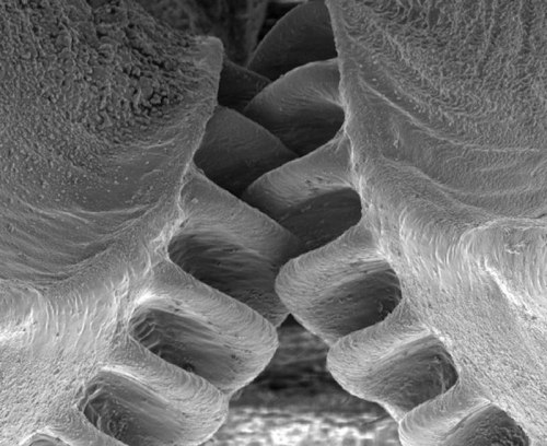
Close-Up of the First Mechanical Gear Ever Found in Nature
The biological form of a mechanical gear was observed in nature for the first time in juvenile planthoppers (Genus: Issus), a common insect that can be found in gardens across Europe.
The insect has hind-leg joints with curved cog-like strips of opposing ‘teeth’ that intermesh, rotating like mechanical gears to synchronize the animal’s legs when it launches into a jump. The finding demonstrates that gear mechanisms previously thought to be solely man-made have an evolutionary precedent.
(Continue Reading)
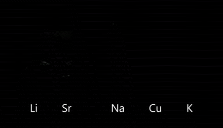
In chemistry, flame tests are used to detect the presence of certain elements, primarily metal ions, by analyzing the colour of the flame given off when heated.
above are Lithium (Li), Strontium (Sr), Sodium (Na), Copper(Cu), and Potassium (K).

Back in the 1960s, the U.S. started vaccinating kids for measles. As expected, children stopped getting measles.
But something else happened.
Childhood deaths from all infectious diseases plummeted. Even deaths from diseases like pneumonia and diarrhea were cut by half.
“So it’s really been a mystery — why do children stop dying at such high rates from all these different infections following introduction of the measles vaccine,” says Michael Mina, a postdoc in biology at Princeton University and a medical student at Emory University.
Scientists Crack A 50-Year-Old Mystery About The Measles Vaccine Photo credit: Photofusion/UIG via Getty Images

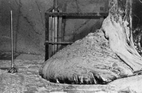
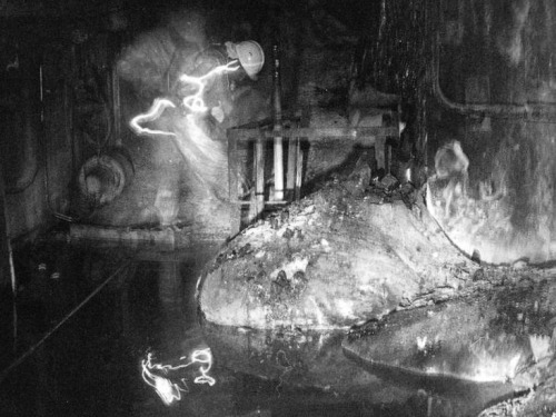
One of the most dangerous pictures ever taken - Elephant’s Foot, Chernobyl. This is a photo of a now dead man next the ‘Elephant’ Foot’ at the Chernobyl power plant.
The image distortions in the photo are created by intense level of radiation almost beyond comprehension. There is no way the person in this photo and the person photographing him could have survived for any more that a few years after being there, even if they quickly ran in, took the photos and ran out again. This photo would be impossible to take today as the rates of radioactive decay are even more extreme now due to a failed military experiment to bomb the reactor core with neuron absorbers. The foot is made up of a small percentage of uranium with the bulk mostly melted sand, concrete and other materials which the molten corium turns into a kind of lava flow. In recent years, it has destroyed a robot which tried to approach it, and the last photos were taken via a mirror mounted to a pole held at the other end of the corridor for a few seconds. It is almost certainly the most dangerous and unstable creation made by humans. These are the effects of exposure: 30 seconds of exposure - dizziness and fatigue a week later 2 minutes of exposure - cells begin to hemorrhage (ruptured blood vessels) 4 minutes - vomiting, diarrhea, and fever 300 seconds - two days to live
-
 pikachusandwich liked this · 2 weeks ago
 -
 igualdadedeexpressao liked this · 3 weeks ago
 -
 howtoleavetheplanet reblogged this · 1 month ago
 -
 hellsiteaffectionate reblogged this · 1 month ago
 -
 librationpoint reblogged this · 3 months ago
 -
 mediumapocalypse reblogged this · 3 months ago
 -
 irrevocablyxo reblogged this · 4 months ago
 -
irrevocablyxo liked this · 4 months ago
-
 no32557038 liked this · 4 months ago
 -
 sherriff-obrien-posting liked this · 4 months ago
 -
 a-song-of-fluff reblogged this · 6 months ago
 -
 buttered-toast-and-sentiment liked this · 6 months ago
 -
 cant-apult reblogged this · 6 months ago
 -
 rahayn reblogged this · 7 months ago
 -
 cravin-paradise liked this · 7 months ago
 -
 azul-ocean24 reblogged this · 7 months ago
 -
 robinzanders liked this · 8 months ago
 -
 sunrisecosmos reblogged this · 8 months ago
 -
 jpyoerixfanxv liked this · 8 months ago
 -
 makesmehappy-tiggs reblogged this · 9 months ago
 -
 crapblog3000 reblogged this · 9 months ago
 -
 messy-complex liked this · 10 months ago
 -
 quantumcellularis reblogged this · 10 months ago
 -
 thatguysbrothersfreind liked this · 10 months ago
 -
 ceilimoose reblogged this · 1 year ago
 -
 wovenlandscape liked this · 1 year ago
 -
 blurred-cat reblogged this · 1 year ago
 -
 aliendesires reblogged this · 1 year ago
 -
blurred-cat liked this · 1 year ago
-
 drachens liked this · 1 year ago
 -
 demonwrestler reblogged this · 1 year ago
 -
demonwrestler liked this · 1 year ago
-
 lith-myathar reblogged this · 1 year ago
 -
lith-myathar liked this · 1 year ago
-
 the-complexity-of-love liked this · 1 year ago
 -
 herald-adaar reblogged this · 1 year ago
 -
 inkstainedgrimm liked this · 1 year ago
 -
 dreamofbecoming reblogged this · 1 year ago
 -
 marshmcore liked this · 1 year ago
 -
 iphigenia-deserves-better liked this · 1 year ago
 -
 cosmos-hime reblogged this · 1 year ago
 -
 mightyflowerwitch reblogged this · 1 year ago
 -
 apprendere reblogged this · 1 year ago
 -
 roamingbadger liked this · 1 year ago
 -
 oldgirlyoungcrone reblogged this · 1 year ago
 -
 runawayfuture reblogged this · 1 year ago
