Science-is-magical - Science Is Magic


More Posts from Science-is-magical and Others

Absence of Serotonin Alters Development and Function of Brain Circuits
Researchers at Case Western Reserve University School of Medicine have created the first complete model to describe the role that serotonin plays in brain development and structure. Serotonin, also called 5-hydroxytryptamine [5-HT], is an important neuromodulator of brain development and the structure and function of neuronal (nerve cell) circuits. The results were published in the current issue of The Journal of Neurophysiology online.
“Our goal in the project was to close the gap in knowledge that exists on role of serotonin in the brain cortex, particularly as it concerns brain circuitry, its electrical activity and function,” said Roberto Fernández Galán, PhD, Assistant Professor in the Department of Neurosciences at Case Western Reserve University School of Medicine. “For the first time, we can provide a complete description of an animal model from genes to behavior—including at the level of neuronal network activity, which has been ignored in most studies to date.”
Dr. Galán and his team used high-density multi-electrode arrays in a mouse model of serotonin deficiency to record and analyze neuronal activity. The study supports the importance of the serotonin which is specified and maintained by a specific gene, the Pet-1 gene – for normal functioning of the neurons, synapses and networks in the cortex, as well as proper development of brain circuitry. Serotonin abnormalities have been linked to autism and epilepsy, depression and anxiety. By more fully elucidating the role of serotonin in the brain, this study may contribute to a better understanding of the development or treatment of these conditions.
“By looking at the circuit level of the brain, we now have new insight into how the brain becomes wired and sensitive to changing serotonin levels.” added Dr. Galán.

Redrawing the brain’s motor map
Neuroscientists at Emory have refined a map showing which parts of the brain are activated during head rotation, resolving a decades-old puzzle. Their findings may help in the study of movement disorders affecting the head and neck, such as cervical dystonia and head tremor.
The results were published in Journal of Neuroscience.
In landmark experiments published in the 1940s and 50s, Canadian neurosurgeon Wilder Penfield and colleagues determined which parts of the motor cortex controlled the movements of which parts of the body.
Penfield stimulated the brain with electricity in patients undergoing epilepsy surgery, and used the results to draw a “motor homunculus”: a distorted representation of the human body within the brain. Penfield assigned control of the neck muscles to a region between those that control the fingers and face, a finding inconsistent with some studies that came later.
Using modern functional MRI (magnetic resonance imaging), researchers at Emory University School of Medicine have shown that the neck’s motor control region in the brain is actually between the shoulders and trunk, a location that more closely matches the arrangement of the body itself.
“We can’t be that hard on Penfield, because the number of cases where he was able to study head movement was quite limited, and studying head motion as he did, by applying an electrode directly to the brain, creates some challenges,” says lead author Buz Jinnah, MD, professor of neurology, human genetics and pediatrics at Emory University School of Medicine.
The new location for the neck muscles makes more sense, because it corresponds to a similar map Penfield established of the sense of touch (the somatosensory cortex), Jinnah says.
Participants in brain imaging studies need to keep their heads still to provide accurate data, so volunteers were asked to perform isometric muscle contraction. They attempted to rotate their heads to the left or the right, even though head movement was restricted by foam padding and restraining straps.
First author Cecilia Prudente, a graduate student in neuroscience who is now a postdoctoral associate at the University of Minnesota, developed the isometric head movement task and obtained internal funding that allowed the study to proceed.
She and Jinnah knew that isometric exercises for the wrist activated the same regions of the motor cortex as wrist movements, and used that as a reference point in their study. During brain imaging, they were able to check that particular muscles were being tensed by directly monitoring volunteers’ muscles electronically.
When volunteers contracted their neck muscles, researchers were able to detect activation in other parts of the brain too, such as the cerebellum and the basal ganglia, which are known to be involved in movement control. This comes as no surprise, Jinnah says, since these regions also control movements of the hands and other body parts.
Prudente, Jinnah and colleagues have conducted a similar study with cervical dystonia patients, with the goal of comparing the patterns of brain activation between healthy volunteers and the patients. Cervical dystonia is a painful condition in which the neck muscles contract involuntarily and the head posture is distorted.
“These results may help guide future studies in humans and animals, as well as medical or surgical interventions for cervical dystonia and other disorders involving abnormal head movements,” Prudente says.


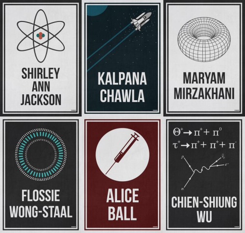
The complete ‘Women Who Changed Science - And The World" collection in honor of the 95th Women’s Equality Day.
Purchase Here!
A puzzling expected value
Pick a (uniformly) random real number from the unit interval [0,1] and repeat this until the sum of all chosen numbers exceeds 1. What is the expected number of real values you will pick?
The quite surprising answer is Eulers constant, e ≈ 2.71828.
A demonstration can be found on Wolfram MathWorld.


What does it take to teach a bee to use tools? A little time, a good teacher and an enticing incentive. Read more here: http://to.pbs.org/2mpRUAz
Credit: O.J. Loukola et al., Science (2017)

-
 proto-indo-european reblogged this · 2 months ago
 -
 mystifyinghatchetfieldtheories liked this · 5 months ago
 -
 saphicspacesociety22 reblogged this · 1 year ago
 -
 nostalgicproteinbag liked this · 1 year ago
 -
 nothing-cereal reblogged this · 1 year ago
 -
 gritinmyslippers reblogged this · 2 years ago
 -
 selendra reblogged this · 2 years ago
 -
 tiphaineaileen liked this · 2 years ago
 -
 misterbatguano reblogged this · 2 years ago
 -
 aleigh75-blog liked this · 2 years ago
 -
 complete360 reblogged this · 2 years ago
 -
 time-to-go-home-blog reblogged this · 2 years ago
 -
time-to-go-home-blog liked this · 2 years ago
-
 loretheuncanny reblogged this · 2 years ago
 -
 whydoesthiskeephappening1121 liked this · 2 years ago
 -
 whereisglory liked this · 2 years ago
 -
 markelba reblogged this · 2 years ago
 -
 cheesewizard liked this · 2 years ago
 -
 followthelion-blog reblogged this · 2 years ago
 -
 duhovipredaka liked this · 2 years ago
 -
 birdsonawyrm liked this · 3 years ago
 -
 da-biggest-fangirl liked this · 3 years ago
 -
 therianimal liked this · 3 years ago
 -
 eclairs-of-emptiness reblogged this · 3 years ago
 -
 random-shit-i-eat reblogged this · 3 years ago
 -
random-shit-i-eat liked this · 3 years ago
-
 pelirroja-peligrosa reblogged this · 3 years ago
 -
 ratswithguns reblogged this · 3 years ago
 -
ratswithguns liked this · 3 years ago
-
 fletcher-bit-me liked this · 3 years ago
 -
 protective-potato reblogged this · 3 years ago
 -
 tittypocalypse reblogged this · 3 years ago
 -
 sassysousa liked this · 3 years ago
 -
 ketsisnotok reblogged this · 3 years ago
 -
ketsisnotok liked this · 3 years ago
-
 x-winging-it reblogged this · 3 years ago
 -
x-winging-it liked this · 3 years ago
-
 dumpst3rgard3n reblogged this · 3 years ago
 -
dumpst3rgard3n liked this · 3 years ago
-
protective-potato liked this · 3 years ago
-
 minecraftfaggot reblogged this · 3 years ago
 -
minecraftfaggot liked this · 3 years ago
-
 deuxiemedunom reblogged this · 3 years ago
