Brain Parasites: Part II.
Brain Parasites: part II.
Taenia solium:

The pork tapeworm, Taenia solium, is the most harmful tapeworm in humans. Taenia solium infection is acquired either from human feces that contains Taenia solium eggs or from uncooked pork which contains larval cysts. If larvae are ingested, they mature into adults in the small intestine. This infection type is called taeniasis and is often asymptomatic. If eggs are ingested, the resulting disease is cysticercosis. It gets its name from larval Taenia solium called cysticercus. Both diseases are common in Africa, Asia, South America and Southern Europe. Taeniasis is rare in Muslim countries since people there do not consume pork.
Keep reading
More Posts from Contradictiontonature and Others

(Image caption: An fMRI scan shows regions of the brain that become active when devoutly religious study participants have a spiritual experience, including a reward center in the brain, the nucleus accumbens. Credit: Jeffrey Anderson)
This is your brain on God
Religious and spiritual experiences activate the brain reward circuits in much the same way as love, sex, gambling, drugs and music, report researchers at the University of Utah School of Medicine. The findings were published in the journal Social Neuroscience.
“We’re just beginning to understand how the brain participates in experiences that believers interpret as spiritual, divine or transcendent,” says senior author and neuroradiologist Jeff Anderson. “In the last few years, brain imaging technologies have matured in ways that are letting us approach questions that have been around for millennia.”
Specifically, the investigators set out to determine which brain networks are involved in representing spiritual feelings in one group, devout Mormons, by creating an environment that triggered participants to “feel the Spirit.” Identifying this feeling of peace and closeness with God in oneself and others is a critically important part of Mormons’ lives — they make decisions based on these feelings; treat them as confirmation of doctrinal principles; and view them as a primary means of communication with the divine.
During fMRI scans, 19 young-adult church members — including seven females and 12 males — performed four tasks in response to content meant to evoke spiritual feelings. The hour-long exam included six minutes of rest; six minutes of audiovisual control (a video detailing their church’s membership statistics); eight minutes of quotations by Mormon and world religious leaders; eight minutes of reading familiar passages from the Book of Mormon; 12 minutes of audiovisual stimuli (church-produced video of family and Biblical scenes, and other religiously evocative content); and another eight minutes of quotations.
During the initial quotations portion of the exam, participants — each a former full-time missionary — were shown a series of quotes, each followed by the question “Are you feeling the spirit?” Participants responded with answers ranging from “not feeling” to “very strongly feeling.”
Researchers collected detailed assessments of the feelings of participants, who, almost universally, reported experiencing the kinds of feelings typical of an intense worship service. They described feelings of peace and physical sensations of warmth. Many were in tears by the end of the scan. In one experiment, participants pushed a button when they felt a peak spiritual feeling while watching church-produced stimuli.
“When our study participants were instructed to think about a savior, about being with their families for eternity, about their heavenly rewards, their brains and bodies physically responded,” says lead author Michael Ferguson, who carried out the study as a bioengineering graduate student at the University of Utah.
Based on fMRI scans, the researchers found that powerful spiritual feelings were reproducibly associated with activation in the nucleus accumbens, a critical brain region for processing reward. Peak activity occurred about 1-3 seconds before participants pushed the button and was replicated in each of the four tasks. As participants were experiencing peak feelings, their hearts beat faster and their breathing deepened.
In addition to the brain’s reward circuits, the researchers found that spiritual feelings were associated with the medial prefrontal cortex, which is a complex brain region that is activated by tasks involving valuation, judgment and moral reasoning. Spiritual feelings also activated brain regions associated with focused attention.
“Religious experience is perhaps the most influential part of how people make decisions that affect all of us, for good and for ill. Understanding what happens in the brain to contribute to those decisions is really important,” says Anderson, noting that we don’t yet know if believers of other religions would respond the same way. Work by others suggests that the brain responds quite differently to meditative and contemplative practices characteristic of some eastern religions, but so far little is known about the neuroscience of western spiritual practices.
The study is the first initiative of the Religious Brain Project, launched by a group of University of Utah researchers in 2014, which aims to understand how the brain operates in people with deep spiritual and religious beliefs.


history meme (french edition) → 7 inventions/achievements (2/7) the first vaccine for rabies by Louis Pasteur & Émile Roux
“Pasteur had, in the early 1880s, a vaccine for rabies, but he was a chemist and not a licensed physician, and potentially liable if he injured or killed a human being. In early july 1885, Joseph Meister, a nine year-old-boy, had been badly mauled and bitten by a rabid dog (…). Pasteur injected young Meister with his rabies vaccine: the boy did not develop rabies and recovered fully from his injuries. Pasteur became a hero, and the Parisian Institue which came to be named in his honour, and of which he was the first director, became the global prototype bacteriological and immunological research institute. By demonstrating beyond doubt that many diseases were transmitted by bacteria and could be prevented from becoming active by pasteurization techniques, Pasteur indeed changed the course of history.” – G. L. Geison, The Private Science of Louis Pasteur.

How the ‘police’ of the cell world deal with 'intruders’ and the 'injured’
The job of policing the microenvironment around our cells is carried out by macrophages. Macrophages are the 'guards’ that patrol most tissues of the body - poised to engulf infections or destroy and repair damaged tissue.
Over the last decade it has been established that macrophages are capable of detecting changes in the microenvironment of human tissues. They can spot pathogen invasion and tissue damage, and mediate inflammatory processes in response, to destroy microbial interlopers and remove and repair damaged tissue. But how do these sentinels of the cell world deal with infection and tissue injury?
Dr Anna Piccinini, an expert in inflammatory signalling pathways in the School of Pharmacy at The University of Nottingham, has discovered that the macrophage’s 'destroy and repair service’ is capable of discriminating between the two distinct threats even deploying a single sensor. As a result, they can orchestrate specific immune responses - passing on information in the form of inflammatory molecules and degrading tissue when they encounter an infection and making and modifying molecular components of the tissue when they detect tissue damage.
Dr Piccinini’s research is published today, Tuesday 30 August 2016, in the academic journal Science Signaling. Her findings could provide future targets for the treatment of diseases with extensive tissue damage such as arthritis or cancer where inflammation plays an increasingly recognized role.
Science Signaling
Macrophage Engulfing Bacteria, Artwork by David Mack


What Happens in the Brain During Unconsciousness?
Researchers are shining a light on the darkness of the unconscious brain. Three new studies add to the body of knowledge.
When patients undergo major surgery, they’re often put under anesthesia to allow the brain to be in an unconscious state.
But what’s happening in the brain during that time?
Three Michigan Medicine researchers are authors on three new articles from the Center for Consciousness Science exploring this question — specifically how brain networks fragment in association with a variety of unconsciousness states.
“These studies come from a long-standing hypothesis my colleagues and I have had regarding the essential characteristic of why we are conscious and how we become unconscious, based on patterns of information transfer in the brain,” says George A. Mashour, M.D., Ph.D., professor of anesthesiology, director of the Center for Consciousness Science and associate dean for clinical and translational research at the University of Michigan Medical School.
In the studies, the team not only explores how the brain networks fragment, but also how better to measure what is happening.
“We’ve been working for a decade to understand in a more refined way how the spatial and temporal aspects of brain function break down during unconsciousness, how we can measure that breakdown and the implications for information processing,” says UnCheol Lee, Ph.D., physicist, assistant professor of anesthesiology and associate director of the Center for Consciousness Science.
Examining different aspects of unconsciousness
The basis for the three studies, as well as other work from the Center for Consciousness Science, comes from a theory Mashour produced during his residency.
“I published a theoretical article when I was a resident in anesthesiology suggesting that anesthesia doesn’t work by turning the brain off, per se, but rather by isolating processes in certain areas of the brain,” Mashour says. “Instead of seeing a highly connected brain network, anesthesia results in an array of islands with isolated cognition and processing. We have taken this thought, as well as the work of others, and built upon it with our research.”
In the study in the Journal of Neuroscience, the team analyzed different areas of the brain during sedation, surgical anesthesia and a vegetative state.
“It’s often suggested that different areas of the brain that typically talk to one another get out of sync during unconsciousness,” says Anthony Hudetz, Ph.D., professor of anesthesiology, scientific director of the Center for Consciousness Science and senior author on the study. “We showed in the early stages of sedation, the information processing timeline gets much longer and local areas of the brain become more tightly connected within themselves. That tightening might lead to the inability to connect with distant areas.”
In the Frontiers in Human Neuroscience study, the team delved into how the brain integrates information and how it can be measured in the real world.
“We took a very complex computational task of measuring information integration in the brain and broke it down into a more manageable task,” says Lee, senior author on the study. “We demonstrated that as the brain gets more modular and has more local conversations, the measure of information integration starts to decrease. Essentially, we looked at how the brain network fragmentation was taking place and how to measure that fragmentation, which gives us the sense of why we lose consciousness.”
Finally, the latest article, in Trends in Neurosciences, aimed to take the team’s previous studies and other work on the subject of unconsciousness and put together a fuller picture.
“We examined unconsciousness across three different conditions: physiological, pharmacological and pathological,” says Mashour, lead author on the study. “We found that during unconsciousness, disrupted connectivity in the brain and greater modularity are creating an environment that is inhospitable to the kind of efficient information transfer that is required for consciousness.”
How these studies can help patients
The team members at the Center for Consciousness Science note that all of this work may help patients in the future.
“We’re looking for a better way to quantify the depth of anesthesia in the operating room and to assess consciousness in someone who has had a stroke or brain damage,” Hudetz says. “For example, we may assume that a patient is fully unconscious based on behavior, but in some cases consciousness can persist despite unresponsiveness.”
The team hopes this and future research could lead to therapeutic strategies for patients.
“We want to understand the communication breakdown that occurs in the brain during unconsciousness so we can precisely target or monitor these circuits to achieve safer anesthesia and restore these circuits to improve outcomes of coma,” Mashour says.
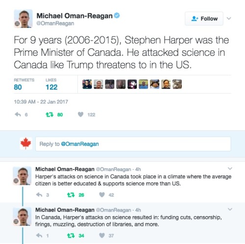


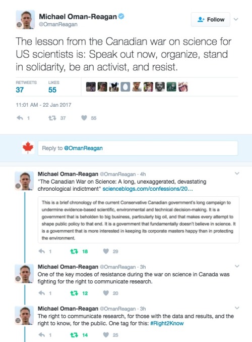
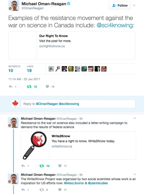

A required read from Michael Oman-Reagan.
This is all true. This all happened in Canada, and its very likely it will happen in the USA under Trump and be worse than Harper’s crackdown on Science ever was.
Links cited in this twitter essay:
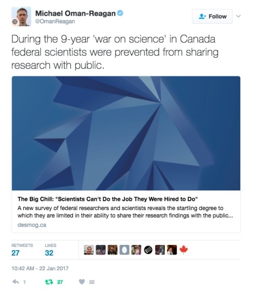
The Big Chill: “Scientists Can’t Do the Job They Were Hired to Do”
More than 1000 Jobs Lost, Climate Program Hit Hard in Coming Environment Canada Cuts
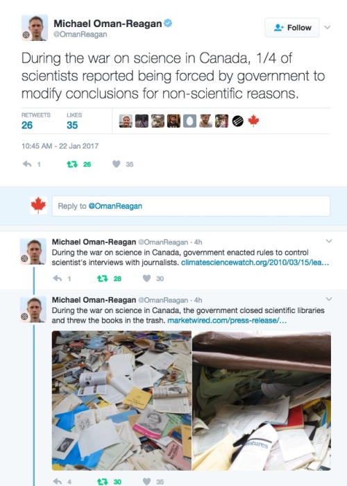
Harper Government Trashes Another Federal Science Library
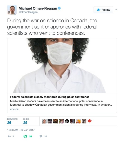
Federal scientists closely monitored during polar conference
Science Silenced: US Scientist Caught in Canadian Muzzle Climate-change scientists feel ‘muzzled’ by Ottawa: Documents
The Canadian War on Science: A long, unexaggerated, devastating chronological indictment
http://ourrighttoknow.ca/
http://write2know.ca/
https://evidencefordemocracy.ca

After the news of an accident in a French drug trial on Friday, you might be wondering what drug trials entail. Here’s a summary sheet on the drug discovery process to clear things up! http://wp.me/p4aPLT-1EZ

The first of the 2016 science Nobel Prizes was announced earlier today. The prize for Medicine or Physiology was awarded to Yoshinori Ohsumi for his work on the mechanisms behind autophagy. Find out more about his Nobel-winning work with this graphic! http://bit.ly/NobelSci2016
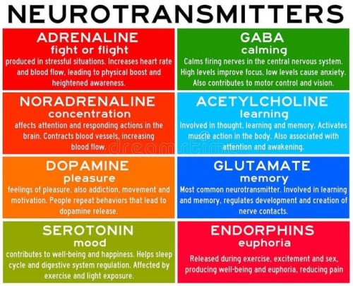
Neurotransmitters are chemicals that help in transmitting signals across a synapse. Different neurotransmitters are associated with different functions. Knowledge about these helps us to treat various neurological conditions by either stimulating or inhibiting these production. #neurology #neuroscience #psychiatry #medicine #medstudynotes #medschool #mbbs #unimed #brain #nervoussystem #physiology #medblog #medblr #medstudent https://www.instagram.com/p/BrM4ocsBqJe/?utm_source=ig_tumblr_share&igshid=12tojib83c32d

A mighty membrane that twists and turns through the gut is starting the new year with a new classification: the structure, called the mesentery, has been upgraded to an organ.
Scientists have known about the structure, which connects a person’s small and large intestines to the abdominal wall and anchors them in place, according to the Mayo Clinic. However, until now, it was thought of as a number of distinct membranes by most scientists. Interestingly, in one of its earliest descriptions, none other than Leonardo da Vinci identified the membranes as a single structure, according to a recent review.
-
 polybrattycrisis liked this · 1 year ago
 -
 glutiamentous liked this · 2 years ago
 -
 kittybluntzz liked this · 3 years ago
 -
 siphonophoraeee liked this · 3 years ago
 -
siphonophoraeee reblogged this · 3 years ago
-
 artificialcherryflavour liked this · 6 years ago
 -
 mileysyrups liked this · 6 years ago
 -
 nospheratusblack liked this · 6 years ago
 -
 brain-bleed liked this · 7 years ago
 -
 nimireshi reblogged this · 7 years ago
 -
 hashtongue reblogged this · 7 years ago
 -
 basementvixen liked this · 7 years ago
 -
 madameliliana1001 liked this · 7 years ago
 -
 optimist-surrounded-by-pessimism liked this · 7 years ago
 -
 sonofthorns-blog liked this · 7 years ago
 -
 thekweenoftheeyesores liked this · 7 years ago
 -
 mayhemandstuff reblogged this · 7 years ago
 -
mayhemandstuff liked this · 7 years ago
-
 alexisyb liked this · 7 years ago
 -
 homotology liked this · 7 years ago
 -
 hopecoloredcrayons liked this · 7 years ago
 -
 belov3d reblogged this · 7 years ago
 -
belov3d liked this · 7 years ago
-
 ravenclawstatus reblogged this · 7 years ago
 -
 mdchicano reblogged this · 7 years ago
 -
 smallandsurviving liked this · 7 years ago
 -
 studybiologyis liked this · 7 years ago
 -
 flyonthewallmedstudent reblogged this · 7 years ago
 -
 ashlarvae reblogged this · 8 years ago
 -
ashlarvae liked this · 8 years ago
-
 bebsylon liked this · 8 years ago
 -
 beccalou890 liked this · 8 years ago
 -
 randomish-things reblogged this · 8 years ago
 -
 hangrypa reblogged this · 8 years ago
 -
 murphy-prinzip liked this · 8 years ago
 -
 valiumcaviar liked this · 8 years ago
 -
 desperately-needs-inspiration reblogged this · 8 years ago
 -
 34ther reblogged this · 8 years ago
 -
shieldmaiden98-blog liked this · 8 years ago
-
 whatshouldwecallmedlab reblogged this · 8 years ago
 -
 pikuuukip reblogged this · 8 years ago
 -
 pikuotaku liked this · 8 years ago
A pharmacist and a little science sideblog. "Knowledge belongs to humanity, and is the torch which illuminates the world." - Louis Pasteur
215 posts