Autonomic Nervous System

Autonomic nervous system
Structure and Function of the Sympathetic and Parasympathetic nervous system
The main function of the autonomic nervous system (ANS) is to assist the body in maintaining a relatively constant internal environment. For example, a sudden increase in systemic blood pressure activates the baroreceptors (those are receptors that detect physical pressure) which in turn modify the activity of the ANS so that the blood pressure is restored to its previous level [1].
The ANS is often regarded as a part of the motor system and is responsible for involuntary action and its effector organs are smooth muscle, cardiac muscle and glands. Another system, the somatic (meaning around the body) nervous system, is responsible for voluntary action in which skeletal muscle is the effector.
The ANS can further be divided into 3 parts: sympathetic, parasympathetic and enteric nervous systems [1][2], with the enteric nervous system sometimes being considered a separate entity [2]. Both parasympathetic and sympathetic nervous systems coexist and work in opposition with each other, ultimately maintaining the correct balance; the activity of one being more active depending on the situation. In a normal resting human, the parasympathetic nervous system dominates, while in a tense and stressful situation, the sympathetic nervous system switches to become dominant.

Figure 1. Structure and function of the central nervous system
This article will be focused on sympathetic and parasympathetic activity from the perspective of:
Anatomy
Biochemical
The sympathetic division provides your “fight or flight” whereas the parasympathetic division helps you to “rest and digest”
Anatomy
Higher centers that control autonomic function include the pons, medulla oblongata and hypothalamus [3].
The pons contains the micturition (urination) and respiratory center.
The medulla oblongata contains the respiratory, cardiac, vomiting, vasomotor and vasodilator centres [4].
The hypothalamus contains the highest concentration of autonomic centres [4]. It contains several centres that control autonomic activities, including heat loss, heat production and conservation, feeding and satiety, as well as fluid intake [4].

Figure 2. Locations of the autonomic control centres of the brain
All 3 structures receive input from certain sources by stimulation of nerve fibres resulting from chemical changes in blood composition like blood pH, blood glucose level, blood osmolarity and volume [4]. Notably, the hypothalamus receives input from cerebral cortex and the limbic system, a system that helps control emotional behaviour [3].
Autonomic promoter neurons are neurons that are found in the brain stem, hypothalamus or even cerebral hemispheres that project to preganglionic neurons (discussed below), where they form synapses with these neurons (5). Hence, input from the higher centres can be relayed to the motor neurons (preganglionic and then postganglionic neurons) which subsequently innervate different body tissues. Changes in the input from these centres could result in responses in those tissues.
The primary functional unit of the sympathetic and parasympathetic nervous system consists of a 2 neuron motor pathway (Figure 3), containing a preganglionic and postganglionic neurons, arranged in series.(2) The two synapse in peripheral ganglion. This clearly distinguishes autonomic motor nervous system and somatic nervous system. The somatic nervous system project from the CNS directly to innervated tissue without any intervening ganglia.(6)

Figure 3. Diagram showing the primary functional unit of the ANS
Sympathetic nervous system
Sympathetic preganglionic neurons mainly are concentrated in the lateral horn in the thoracic (T1-12) and upper lumbar (L1 &2) segments of the spinal cord (Figure 4).
The preganglionic axons leave the spinal cord in 3 ways:
Through the paravertebral ganglion
The preganglionic axon may synapse with postganglionic neurons in this ganglion or some axon may travel rostrally or caudally within the sympathetic trunk before forming synapse with a postganglionic neurons in a different paravertebral ganglion.
Through the prevertebral ganglion
Some preganglionic axons pass the paravertebral ganglion (no synapse occur) and form synapse with postganglionic neurons in prevertebral ganglion, also known as collateral ganglion.
Directly to the organs without any synapse
Some preganglionic axons pass through the sympathetic trunk (no synapse) and end directly on cells of the adrenal medulla, which are equivalent to postganglionic cell.
Parasympathetic nervous system
The parasympathetic preganglionic neurons are located in several cranial nerve nuclei in the brain stem and some are found in the S3 and S4 segments of the sacral spinal cord (Figure 4). The parasympathetic postganglionic neurons are located in cranial ganglia, including the ciliary ganglion, the pterygopalatine, submandibular ganglia, and the otic ganglion. Other ganglia are present near or in the walls of visceral organs. Similarly, the preganglionic neurons form synapse with the postganglionic neurons in the ganglia.

Figure 4. Anatomy of the ANS and how its nuerons innervate tissues
After knowing how nerves connect from the CNS to PNS and to different organs, we will now consider some of the neurotransmitters that are being released at different nerve terminals. It is the binding of these neurotransmitters to the receptors on the effectors that leads to biochemical and physiological changes. Some of the neurotransmitters in use are:
For the synapse between pre and postganglionic neurons mentioned above, the neurotransmitter that is released by the preganglionic axon terminal, is acetylcholine. The corresponding receptors are found on the postsynaptic membrane of postganglionic nerves and are nicotinic receptors.
Parasympathetic postganglionic nerve terminals also release acetylcholine.
Sympathetic postganglionic nerve terminals release mostly noradrenaline
The adrenal medulla receives direct stimulation from sympathetic preganglionic innervation, releases mainly adrenaline (80%) and some noradrenaline into the blood stream. In this case, both adrenaline and noradrenaline act as hormones as they are transported via blood circulating system to target organs instead of neuronal pathway.
Strangely, for the sympathetic postganglionic nerves that innervate the sweat glands, the nerves release acetylcholine (normally only by parasympathetic postganglionic nerve) instead.
1. H.P.Rang, J.M.Ritter, R.J.Flower GH. RANG & DALE’S Pharmacology. In: 8th ed. ELSEVIER CHURCHILL LIVINGSTONE; 2016. p. 145.
2. Bruce M. Koeppen BAS. BERNE & LEVY PHYSIOLOGY. In: 6th ed. MOSBY ELSEVIER; 2010. p. 218.
3. Cholinergic transmission [Internet]. 2015. Available from: http://www.dartmouth.edu/~rpsmith/Cholinergic_Transmission.html
4. Bruce M. Koeppen BAS. BERNE & LEVY PHYSIOLOGY. In: 6th ed. MOSBY ELSEVIER; 2016. p. 44.
More Posts from Contradictiontonature and Others
Meet the obscure microbe that influences climate, ocean ecosystems, and perhaps even evolution
Penny Chisholm has had a 35-year love affair—with a microbe. For her, it’s been the perfect partner—elusive during courting, a source of intellectual fulfillment, and still full of mystery decades after their introduction during an ocean cruise.
To look at, the object of her passion is just a green mote, floating in vast numbers in the world’s oceans. But Chisholm has found hidden complexity within Prochlorococcus, a cyanobacterium that is the smallest, most abundant photosynthesizing cell in the ocean—responsible for 5% of global photosynthesis, by some estimates. Its many different versions, or ecotypes, thrive from the sunlit sea surface to a depth of 200 meters, where light is minimal. Collectively the “species” boasts an estimated 80,000 genes—four times what humans have, and plenty to deal with whatever the world’s oceans throw at it. “It’s a beautiful little life machine and like a superorganism,” Chisholm says. “It’s got a story to tell us.”

Quote by #rosalindfranklin How do you make science a part of your life? What are you doing to fight for scientific literacy? More quotes and questions in my #ilovescience journal. #womeninscience #scientificliteracy


Leopard shark makes world-first switch from sexual to asexual reproduction
A leopard shark in an Australian aquarium has reproduced asexually after being separated from her mate.
It is the first reported case of a shark switching from sexual to asexual or parthenogenetic reproduction and only the third reported case among all vertebrate species.
The leopard shark, Leonie, was captured in the wild in 1999 and introduced to a male shark at the Reef HQ aquarium in Townsville, Queensland, in 2006. Leopard sharks are also known as zebra sharks.
One of the baby sharks born to leopard sharks at Reef HQ aquarium in Townsville, who have produced live young through asexual reproduction. Photograph: Tourism and Events Queensland
Leonie, the world’s first shark known to have switched from sexual to asexual reproduction, at Reef HQ aquarium in Townsville. Photograph: Tourism and Events Queensland
Thorny life of new-born neurons
Even in adult brains, new neurons are generated throughout a lifetime. In a publication in the scientific journal PNAS, a research group led by Goethe University describes plastic changes of adult-born neurons in the hippocampus, a critical region for learning: frequent nerve signals enlarge the spines on neuronal dendrites, which in turn enables contact with the existing neural network.

Practise makes perfect, and constant repetition promotes the ability to remember. Researchers have been aware for some time that repeated electrical stimulation strengthens neuron connections (synapses) in the brain. It is similar to the way a frequently used trail gradually widens into a path. Conversely, if rarely used, synapses can also be removed – for example, when the vocabulary of a foreign language is forgotten after leaving school because it is no longer practised. Researchers designate the ability to change interconnections permanently and as needed as the plasticity of the brain.
Plasticity is especially important in the hippocampus, a primary region associated with long-term memory, in which new neurons are formed throughout life. The research groups led by Dr Stephan Schwarzacher (Goethe University), Professor Peter Jedlicka (Goethe University and Justus Liebig University in Gießen) and Dr Hermann Cuntz (FIAS, Frankfurt) therefore studied the long-term plasticity of synapses in new-born hippocampal granule cells. Synaptic interconnections between neurons are predominantly anchored on small thorny protrusions on the dendrites called spines. The dendrites of most neurons are covered with these spines, similar to the thorns on a rose stem.
In their recently published work, the scientists were able to demonstrate for the first time that synaptic plasticity in new-born neurons is connected to long-term structural changes in the dendritic spines: repeated electrical stimulation strengthens the synapses by enlarging their spines. A particularly surprising observation was that the overall size and number of spines did not change: when the stimulation strengthened a group of synapses, and their dendritic spines enlarged, a different group of synapses that were not being stimulated simultaneously became weaker and their dendritic spines shrank.
“This observation was only technically possible because our students Tassilo Jungenitz and Marcel Beining succeeded for the first time in examining plastic changes in stimulated and non-stimulated dendritic spines within individual new-born cells using 2-photon microscopy and viral labelling,” says Stephan Schwarzacher from the Institute for Anatomy at the University Hospital Frankfurt. Peter Jedlicka adds: “The enlargement of stimulated synapses and the shrinking of non-stimulated synapses was at equilibrium. Our computer models predict that this is important for maintaining neuron activity and ensuring their survival.”
The scientists now want to study the impenetrable, spiny forest of new-born neuron dendrites in detail. They hope to better understand how the equilibrated changes in dendritic spines and their synapses contribute the efficient storing of information and consequently to learning processes in the hippocampus.
Toxic Alzheimer’s Protein Spreads Through Brain Via Extracellular Space
A toxic Alzheimer’s protein can spread through the brain—jumping from one neuron to another—via the extracellular space that surrounds the brain’s neurons, suggests new research from Karen Duff, PhD, and colleagues at Columbia University Medical Center.

(Image caption: Orange indicates where tau protein has traveled from one neuron to another. Credit: Laboratory of Karen Duff, PhD)
The spread of the protein, called tau, may explain why only one area of the brain is affected in the early stages of Alzheimer’s but multiple areas are affected in later stages of the disease.
“By learning how tau spreads, we may be able to stop it from jumping from neuron to neuron,” says Dr. Duff. “This would prevent the disease from spreading to other regions of the brain, which is associated with more severe dementia.”
The idea the Alzheimer’s can spread through the brain first gained support a few years ago when Duff and other Columbia researchers discovered that tau spread from neuron to neuron through the brains of mice.
In the new study, lead scientist Jessica Wu, PhD, of the Taub Institute discovered how tau travels by tracking the movement of tau from one neuron to another. Tau, she found, can be released by neurons into extracellular space, where it can be picked up by other neurons. Because tau can travel long distances within the neuron before its release, it can seed other regions of the brain.
“This finding has important clinical implications,” explains Dr. Duff. “When tau is released into the extracellular space, it would be much easier to target the protein with therapeutic agents, such as antibodies, than if it had remained in the neuron.”
A second interesting feature of the study is the observation that the spread of tau accelerates when the neurons are more active. Two team members, Abid Hussaini, PhD, and Gustavo Rodriguez, PhD, showed that stimulating the activity of neurons accelerated the spread of tau through the brain of mice and led to more neurodegeneration.
Although more work is needed to examine whether those findings are relevant for people, “they suggest that clinical trials testing treatments that increase brain activity, such as deep brain stimulation, should be monitored carefully in people with neurodegenerative diseases,” Dr. Duff says.


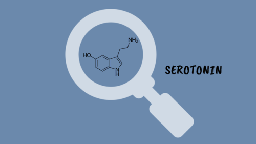
How Do Antidepressants Work? (Video)
Your brain is a network of billions of neurones, all somehow connected to each other. At this very second, millions of impulses are being transmitted through these connections carrying information about what you can see and hear, as well as your emotional state. It’s an incredibly complex system but sometimes things go wrong. Despite extensive research, we are still not certain on the biology that underlies mental illnesses- including depression. However, we have come pretty far in developing effective treatments.

It’s a textbook moment centuries in the making: more than 200 years after scientists started investigating how water molecules conduct electricity, a team has finally witnessed it happening first-hand.
It’s no surprise that most naturally ocurring water conducts electricity incredibly well - that’s a fact most of us have been taught since primary school. But despite how fundamental the process is, no one had been able to figure out how it actually happens on the atomic level.
“This fundamental process in chemistry and biology has eluded a firm explanation,” said one of the team, Anne McCoy from the University of Washington. “And now we have the missing piece that gives us the bigger picture: how protons essentially ‘move’ through water.”
Continue Reading.

It’s a tremendous Trilobite Tuesday!
When most of us think about trilobites, we imagine rather small creatures that inhabited the ancient seas. Indeed, most members of the more than 25,000 scientifically recognized trilobite species were less that three inches in length. Occasionally, however, paleontologists encounter a megafauna where, due to a variety of circumstances, the trilobite species were huge. One of these megafaunas can be found near the small Portuguese town of Arouca where the 450 million year-old Valongo formation produces prodigious numbers of exceptionally large Ordovician-age trilobites, such as this 41 cm Hungioides bohemicus. Other trilobite magafaunas appear sporadically around the globe, including Cambrian locations in Morocco and Devonian outcrops in Nevada.
Meet many more trilobites on the Museum website.

Hepatitis B in a Laboratory!! #serology #infection #liver #hepatitis #medicine #medstudent #medstudynotes #virus #medschool #microbiology #pathology https://www.instagram.com/p/BrIYa-LheyH/?utm_source=ig_tumblr_share&igshid=lqm0j92yobw0
Last Week In Science

1. Broad Institute wins CRISPR patent battle
basically UC Berkely has rights to use CRISPR in “all kinds of cells” and Broad has rights in “eukaryotic cells” (yay legal system). Anticipate more legal battles since there are more types of CRISPR techniques
2. Human genome editing gets the OK to prevent “serious heritable diseases and conditions only”
Bioshock likely to happen in 50 years as “serious disease” dwindles in to “mediocre disease” and finally “what the hell let’s shoot fire from our hands”

3. With the EPA at risk of being destroyed, what was life like before the EPA?
4. Congress wants to shift Earth Science away from NASA (and focus on deep space)
4.1 Coders continue to save climate data
5. This years winners of underwater photos

6. Got trash on your power lines? That’s alright just attach a flamethrower to a drone, no worries
7. Fungicides bring us closer to figuring out why all of the bees are dying
7.1 (but who cares right? we can just make quadcopters do all the work)
8. Australia is HOT AS BALLS

9. Aztecs probably died off from salmonella outbreak
10. Our genetic past and present sanitary world lead to increased autoimmunity and allergy
10.1 Getting the right microbiome early on is so important for health
11. New Zealand on a new continent might make maps include it more often

12. Now you realize how slow the speed of light is on a cosmic scale
13. Meta-Analysis shows Vitamin D supplementation provides “modest protective effect” from respiratory infections like the flu or cold
14. Watch Yosemite’s Horsetail and its annual “FireFall” (image via Robert Minor)

15. Trump’s press conference makes people wonder if he is mentally ill and if we should start testing old ass presidents for dementia
16. He continue’s to spew more anti-vaccine bullshit, showing his ignorance of science and RFK Jr.’s scam needs “just one study” to change his mind
16.1 more than 350 organizations write to Trump to assure his feeble mind that vaccines are safe
17. Simple fractal patterns are key to Rorschach test

18. Imagine shining a light somewhere on your body and microscopic bots deliver drugs there
19. How flat can a planet be?
20. Triangulene created for the first time
Who needs carefully planned chemical reactions when you can just blast hydrogens off with electricity?

21. All of the nerdy. Valentine’s. you. will. ever. need.
22. Help find Planet 9 in your spare time
22.1 Don’t have time? then do science while your computer is idle!

-
 cosmixoracle liked this · 6 years ago
 -
 perv--deartmallory liked this · 7 years ago
 -
 laurenwilzo97-blog liked this · 8 years ago
 -
 migg0705-blog liked this · 8 years ago
 -
 pre-med-disaster-blog reblogged this · 8 years ago
 -
 pre-med-disaster-blog liked this · 8 years ago
 -
 funny-ja liked this · 8 years ago
 -
 harrysfavouritecat reblogged this · 8 years ago
 -
 wavesofgoodenergy liked this · 8 years ago
 -
 harrysfavouritecat liked this · 8 years ago
 -
 harrysfavouritecat reblogged this · 8 years ago
 -
 jgriffey reblogged this · 8 years ago
 -
 jgriffey liked this · 8 years ago
 -
 justacuriouswanderer liked this · 8 years ago
 -
 monserrathsc reblogged this · 8 years ago
 -
 imlostinstereo liked this · 8 years ago
 -
 healthygirlbehavior liked this · 8 years ago
 -
 pheobewankenobi reblogged this · 8 years ago
 -
 pheobewankenobi liked this · 8 years ago
 -
 yolandaarchi liked this · 8 years ago
 -
 restlessss liked this · 8 years ago
 -
 camillethewiz liked this · 8 years ago
 -
 jenanne-nuhaily reblogged this · 8 years ago
 -
 thanks-4 liked this · 8 years ago
 -
 specialagentparrish liked this · 8 years ago
 -
 backseatbucky reblogged this · 8 years ago
 -
 backseatbucky liked this · 8 years ago
 -
 chenstead reblogged this · 8 years ago
 -
 chenstead liked this · 8 years ago
 -
 rabolas liked this · 8 years ago
 -
 tljohnson1988 liked this · 8 years ago
 -
 randomtodaymaybe reblogged this · 8 years ago
 -
 sobusytolive liked this · 9 years ago
 -
 saucererr reblogged this · 9 years ago
 -
 09102016 liked this · 9 years ago
 -
 eurekareference reblogged this · 9 years ago
 -
 let-it-be-tome liked this · 9 years ago
 -
 carmentheninja liked this · 9 years ago
 -
 lostandfaceless reblogged this · 9 years ago
 -
 tilbnifpoe reblogged this · 9 years ago
A pharmacist and a little science sideblog. "Knowledge belongs to humanity, and is the torch which illuminates the world." - Louis Pasteur
215 posts