Let’s Hop This Simple Solution Sticks Around (x) | Follow @the-future-now


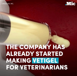


Let’s hop this simple solution sticks around (x) | follow @the-future-now
More Posts from Science-is-magical and Others


A human brain has around 86 billion neurons, and the communication between these neurons are constant. The sheer scale of these interactions mean a computer (an EEG) can register this electrical activity, with different frequencies indicating different mental states.
Sources

‘Smart fat cells’ cross blood-brain barrier to catch early brain tumors
An MRI contrast agent that can pass through the blood-brain barrier will allow doctors to detect deadly brain tumors called gliomas earlier, say Penn State College of Medicine researchers. This ability opens the door to make this fatal cancer treatable.
Gliomas are brain tumors that arise from glial cells, which help nerve cells to stay connected and send signals throughout the body.
Cancerous gliomas are uniformly fatal, with a median survival rate of 14 months from the time of diagnosis. But a new nanotechnology approach developed by Xiaoli Liu and Madhan Kumar in the Department of Neurosurgery could transform gliomas from a death sentence into a treatable condition.
Patients diagnosed with a malignant glioma can undergo surgery, chemotherapy and radiation to destroy the tumor, but the cancer will return.
“Patients typically don’t die from the tumor they initially presented with. Rather, they die from new tumors that come back in other parts of the brain,” said James Connor, Distinguished Professor of Neurosurgery.
These new gliomas tend to grow quickly and are often resistant to treatment because they spring from cancer cells that survived the first therapeutic assault. Glioma patients have follow-up MRIs to detect new brain cancers but the tests do not catch the tumors early enough to save lives.
That is because contrast agents used to outline gliomas on an MRI can only pass the protective blood-brain barrier once the tumors have grown large enough to cause damage to the barrier. Until then, the blood-brain barrier blocks 98 percent of small molecules and all large molecules from entering the brain.
To overcome this deadly limitation, Penn State researchers created “smart fat cells” called liposomes that can pass the blood-brain barrier in mice, seek out tiny cancerous gliomas like heat-seeking missiles and light them up on an MRI. The liposomes are loaded with the most commonly used contrast agent, Magnevist. On their surface, the liposomes are studded with proteins that target receptors on glioma cells.
The new contrast agent delivery system is more sensitive than traditional contrast-enhanced MRI, Connor said.
The researchers found that the liposomes entered the brain in healthy mice with uncompromised blood-brain barriers. Both the conventional and the new technique found large gliomas in mice with cancer, but only the liposome-encapsulated agent was able to detect smaller early-stage tumors. “The goal is to be able to get down to detecting single cancer cells,” Connor said.
The study was published in Journal of Neuro-Oncology.
It is not exactly known how the liposomes get past the intact blood-brain barrier, but they apparently do it without causing damage. In the study, mice showed no harm from the treatment.
This novel approach is an alternative to ultrasound, another promising method researchers are studying to get therapeutic agents into the brain. Ultrasound, however, causes temporary disruption to the blood-brain barrier, which allows not only the therapeutic agent to enter the brain, but also blood which could have medical implications.
“Ultrasound, with all of its good qualities, is disruptive to the blood-brain barrier, whereas we can get an agent to cross it without causing disruption.” Connor said.
The researchers said that in the future, smart fat cells will deliver chemotherapeutic drugs, along with contrast agents, to brain tumor patients so that cancer cells can be detected and wiped out in one step. They recently presented research on these next-generation liposomes at the Society for Neuro-Oncology meeting in San Antonio.
So they found this adorable little dinosaur called Anchiornis


See those feathers? The skeleton they found was so well-preserved that scientists were able to examine the pigment cells in the feathers and compare them to those of modern day birds.
And they were able to do this with such accuracy that they know the coloration of this dinosaur. In life it looked something like this.

It just baffles me that we know the color patterns of an animal that has been dead for 161 million years

Slime mold was grown on an agar gel plate shaped like America and food sources were placed where America’s large cities are.
The result? A possible look at how to best build public transportation.
I just really like the idea of slime mold on a map of the US. It’s beautiful.
A puzzling expected value
Pick a (uniformly) random real number from the unit interval [0,1] and repeat this until the sum of all chosen numbers exceeds 1. What is the expected number of real values you will pick?
The quite surprising answer is Eulers constant, e ≈ 2.71828.
A demonstration can be found on Wolfram MathWorld.


ALL ROLLED UP
A newly identified mineral christened merelaniite tightly rolls up like a scroll as it crystallizes, forming shiny dark gray needles up to a few millimeters in length (Minerals 2016, DOI: 10.3390/min6040115). The overall formula of the mineral is Mo₄Pb₄VSbS₁₅. It crystallizes into a sheet composed primarily of alternating ultrathin layers of MoS₂ and PbS. “It’s like a natural nanocomposite,” says research team leader John A. Jaszczak of Michigan Technological University. Strain from the interacting layers likely causes the crystalline sheets to wrap around themselves as they grow. Jaszczak and coworkers named the mineral for the Merelani mining district in Tanzania, where the merelaniite samples originated. Collaborating research institutions included the U.K. Natural History Museum, U.S. National Museum of Natural History, and University of Florence.
Credit: Minerals (both)
Related C&EN content:
Minerals in Medicine Exhibition
Worldwide Hunt For Missing Carbon Minerals Begins

-
 dragonfly1132861 liked this · 7 months ago
 -
 dragondusk-moved liked this · 1 year ago
 -
 quarkyw0lf13 liked this · 1 year ago
 -
 i-fear-neither-death-nor-pain reblogged this · 1 year ago
 -
 thiefinwhite liked this · 1 year ago
 -
 sarahs-shadow reblogged this · 1 year ago
 -
 pencil-drawn-protagonist liked this · 1 year ago
 -
 yukinohananana017 reblogged this · 2 years ago
 -
yukinohananana017 liked this · 2 years ago
-
 roundaboutnow liked this · 2 years ago
 -
 stvoyejvintovkoj liked this · 2 years ago
 -
 notadragon liked this · 2 years ago
 -
 dvinicius2 liked this · 2 years ago
 -
 fuinjustsu liked this · 2 years ago
 -
 purple-platypus liked this · 2 years ago
 -
 fantasticflynn reblogged this · 3 years ago
 -
fantasticflynn liked this · 3 years ago
-
 filez34 liked this · 3 years ago
 -
 thehoneymushroomhealer liked this · 3 years ago
 -
 t-c-art-inspiration reblogged this · 3 years ago
 -
 terrific-togekiss reblogged this · 3 years ago
 -
 daughterofgaia liked this · 3 years ago
 -
 lollo12589 liked this · 3 years ago
 -
 thedragonreborn24 liked this · 3 years ago
 -
 the-real-kilgore-trout liked this · 3 years ago
 -
 oroichonno liked this · 3 years ago
 -
 ratlicker84zero liked this · 3 years ago
 -
 akjzsd reblogged this · 3 years ago
 -
 chuck-phuckett reblogged this · 3 years ago
 -
 the-monster-mash liked this · 3 years ago
 -
 phoenix0515 liked this · 3 years ago
 -
 faded-coat-of-blue liked this · 3 years ago
 -
 ardashiri reblogged this · 3 years ago
 -
ardashiri liked this · 3 years ago
-
 ichithecupcake reblogged this · 3 years ago
 -
ichithecupcake liked this · 3 years ago
-
 thessalian reblogged this · 3 years ago
 -
 omni-too reblogged this · 3 years ago
 -
 helloturtlebutter liked this · 4 years ago
 -
 nottoofictional liked this · 4 years ago
 -
 sekto78 liked this · 4 years ago
 -
 talon24 reblogged this · 4 years ago
 -
 defaultjane reblogged this · 4 years ago
 -
 peachy-jeans liked this · 4 years ago
