An International Team Of Researchers Has Finally Decoded The Science Behind A Plant Responsible For No

An international team of researchers has finally decoded the science behind a plant responsible for no small degree of human misery: poison ivy.
For the first time, we now know why poison ivy leaves – the bane of campers, hikers, and overly curious kids alike – make us itch, and the answer lies in a key molecule called CD1a, which scientists have long known about but didn’t fully understand until now.
“For over 35 years we have known CD1a is abundant in the skin,” says researcher Jerome Le Nours from Monash University in Australia. “Its role in inflammatory skin disorders has been difficult to investigate and until now has been really unclear.”
One of the reasons for that lack of clarity has been that many experiments on skin disorders involve animal testing – specifically lab mice. And mice don’t produce CD1a, effectively creating a kind of ‘blind spot’ in the studies up to this point.
To get around this and examine whether CD1a might play a part in how human skin reacts when we brush up against poison ivy (Toxicodendron radicans) and similar rash-inducing plants, the researchers genetically engineered mice that did produce the molecule.
In doing so, the team found that CD1a – a protein that plays an important role in our immune systems – triggers a skin-based allergic reaction when we come into contact with urushiol, the allergen that functions as the active ingredient in plants like poison ivy, poison oak, and poison sumac.
When urushiol interacts with skin cells called Langerhans cells, the CD1a proteins (which are expressed by Langerhans cells) activate the immune system’s T cells. In turn, the T cells produce two proteins – interleukin 17 and interleukin 22 – which cause inflammation and itchiness.
Continue Reading.
More Posts from Science-is-magical and Others


Redrawing the brain’s motor map
Neuroscientists at Emory have refined a map showing which parts of the brain are activated during head rotation, resolving a decades-old puzzle. Their findings may help in the study of movement disorders affecting the head and neck, such as cervical dystonia and head tremor.
The results were published in Journal of Neuroscience.
In landmark experiments published in the 1940s and 50s, Canadian neurosurgeon Wilder Penfield and colleagues determined which parts of the motor cortex controlled the movements of which parts of the body.
Penfield stimulated the brain with electricity in patients undergoing epilepsy surgery, and used the results to draw a “motor homunculus”: a distorted representation of the human body within the brain. Penfield assigned control of the neck muscles to a region between those that control the fingers and face, a finding inconsistent with some studies that came later.
Using modern functional MRI (magnetic resonance imaging), researchers at Emory University School of Medicine have shown that the neck’s motor control region in the brain is actually between the shoulders and trunk, a location that more closely matches the arrangement of the body itself.
“We can’t be that hard on Penfield, because the number of cases where he was able to study head movement was quite limited, and studying head motion as he did, by applying an electrode directly to the brain, creates some challenges,” says lead author Buz Jinnah, MD, professor of neurology, human genetics and pediatrics at Emory University School of Medicine.
The new location for the neck muscles makes more sense, because it corresponds to a similar map Penfield established of the sense of touch (the somatosensory cortex), Jinnah says.
Participants in brain imaging studies need to keep their heads still to provide accurate data, so volunteers were asked to perform isometric muscle contraction. They attempted to rotate their heads to the left or the right, even though head movement was restricted by foam padding and restraining straps.
First author Cecilia Prudente, a graduate student in neuroscience who is now a postdoctoral associate at the University of Minnesota, developed the isometric head movement task and obtained internal funding that allowed the study to proceed.
She and Jinnah knew that isometric exercises for the wrist activated the same regions of the motor cortex as wrist movements, and used that as a reference point in their study. During brain imaging, they were able to check that particular muscles were being tensed by directly monitoring volunteers’ muscles electronically.
When volunteers contracted their neck muscles, researchers were able to detect activation in other parts of the brain too, such as the cerebellum and the basal ganglia, which are known to be involved in movement control. This comes as no surprise, Jinnah says, since these regions also control movements of the hands and other body parts.
Prudente, Jinnah and colleagues have conducted a similar study with cervical dystonia patients, with the goal of comparing the patterns of brain activation between healthy volunteers and the patients. Cervical dystonia is a painful condition in which the neck muscles contract involuntarily and the head posture is distorted.
“These results may help guide future studies in humans and animals, as well as medical or surgical interventions for cervical dystonia and other disorders involving abnormal head movements,” Prudente says.

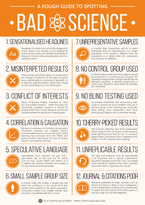
Version 1 of ‘A Rough Guide to Spotting Bad Science’. Thanks for everyone’s suggestions earlier in the week, attempted to include as many of them as possible!
Download link here: http://wp.me/p4aPLT-ap


the drum is filled with hot steam and then sprayed with cold water. the pressure on the outside of the drum is far more than inside. the pressures try to maintain and find balance taking the drum as a casualty.

In chemistry, flame tests are used to detect the presence of certain elements, primarily metal ions, by analyzing the colour of the flame given off when heated.
above are Lithium (Li), Strontium (Sr), Sodium (Na), Copper(Cu), and Potassium (K).

Acting outrageous and making a complete fool of yourself while drunk has been linked to a genetic mutation. It blocks the production of one of the body’s serotonin receptors, which can affect mood swings, impulsive behavior, and decision making. So far, the mutation has only been found in Finnish people, but the discovery is helping researchers understand more about the role serotonin plays in your body. Source Source 2


TYPES OF COLOR-BLINDNESS
1. Normal vision
2. Deuteranopia
3. Tritanopia
4. Monochromacy - An extremely rare type of color-blindness in which sufferers can see only in shades of grey, and perceive no color at all. About 1 in 33,000 people is born with this condition.
(Source)
-
 electricslug reblogged this · 5 years ago
 -
 science-is-magical reblogged this · 8 years ago
 -
 casuallyjollybird reblogged this · 8 years ago
 -
casuallyjollybird liked this · 8 years ago
-
 cedar-glade reblogged this · 8 years ago
 -
 flowerforever reblogged this · 8 years ago
 -
 itsjustmerockyracoon liked this · 8 years ago
 -
 anemonepiperi liked this · 8 years ago
 -
 plantasyotrascosas reblogged this · 8 years ago
 -
 mordekarl liked this · 8 years ago
 -
 saltysalmonella liked this · 8 years ago
 -
 transwhore-blog1 liked this · 8 years ago
 -
 chemistdazzle reblogged this · 8 years ago
 -
 nai21things reblogged this · 8 years ago
 -
nai21things liked this · 8 years ago
-
 defectivebydesign reblogged this · 8 years ago
 -
 peraudacia-adastra reblogged this · 8 years ago
 -
 lunaslashsea reblogged this · 8 years ago
 -
 ganesg-blog liked this · 8 years ago
 -
 justasimplejellyfish liked this · 8 years ago
 -
 trashconnoisseur liked this · 8 years ago
 -
 invisiblesimmer reblogged this · 8 years ago
 -
invisiblesimmer liked this · 8 years ago
-
 albr3relationshipgoals-blog liked this · 8 years ago
 -
 alipaoli liked this · 8 years ago
 -
 chewbaccaaah reblogged this · 8 years ago
 -
 kacishmaci-blog liked this · 8 years ago
 -
 liobits reblogged this · 8 years ago
 -
 devourer-of-acetone reblogged this · 8 years ago
 -
 letsmaketroubleinthedreamworld liked this · 8 years ago
 -
 boreal-sea reblogged this · 8 years ago
 -
boreal-sea liked this · 8 years ago
-
 iyeshaisriley reblogged this · 8 years ago
 -
 thesillyhaley reblogged this · 8 years ago
 -
 howlowcanrubisco-blog reblogged this · 8 years ago
 -
 pastellieria reblogged this · 8 years ago
 -
 annularlight reblogged this · 8 years ago
 -
annularlight liked this · 8 years ago
-
 wrenstep reblogged this · 8 years ago
 -
wrenstep liked this · 8 years ago
-
 adventuresoftheawkward liked this · 8 years ago
 -
 singingstranger liked this · 8 years ago
 -
 thatoneambigousgirl liked this · 8 years ago
 -
 wndr-wll reblogged this · 8 years ago
