Why Number Are The Way They Are

Why Number Are The Way They Are
More Posts from Science-is-magical and Others

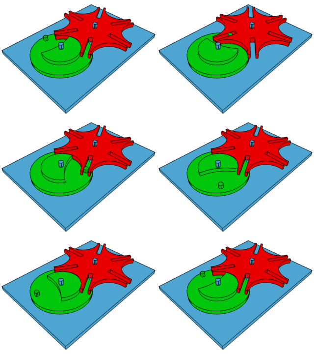
How the Geneva Drive (the mechanical step that makes the second hand on a clock work by turning constant rotation into intermittent motion) works.

There’s Evidence of a New Ninth Planet. For real!
Caltech researchers have found evidence of a giant planet tracing a bizarre, highly elongated orbit in the outer solar system. The object, nicknamed Planet Nine, has a mass about 10 times that of Earth and orbits about 20 times farther from the sun on average than does Neptune, farthest planet from the Sun. In fact, it would take this new planet between 10,000 and 20,000 years to make just one full orbit around the sun.
Planetary scientists, Konstantin Batygin and Mike Brown, describe their work in the current issue of the Astronomical Journal and show how Planet Nine helps explain a number of mysterious features of the field of icy objects and debris beyond Neptune known as the Kuiper Belt.
Unlike the class of smaller objects now known as dwarf planets, Planet Nine gravitationally dominates its neighborhood of the solar system. In fact, it dominates a region larger than any of the other known planets.
Batygin and Brown predicted the planet’s existence through mathematical modeling and computer simulations but have not yet observed the object directly.
To put it briefly, Batygin and Brown inferred its presence from the peculiar clustering of six previously known objects that orbit beyond Neptune. They say there’s only a 0.007% chance that the clustering could be a coincidence. Instead, they say, a planet has shepherded the six objects into their strange elliptical orbits, tilted out of the plane of the solar system. It wasn’t the first possibility they investigated and they ran different simulations until finding that an anti-aligned orbit of the ninth planet prevents the Kuiper Belt objects from colliding with it and keeps them aligned. read more here
Diagram: The six most distant known objects in the solar system with orbits beyond Neptune (magenta) all mysteriously line up in a single direction. Also, when viewed in three dimensions, they all tilt nearly identically away from the plane of the solar system. A planet with in a distant eccentric orbit anti-aligned with the other six objects (orange) is required to maintain this configuration. The diagram was created using WorldWide Telescope. Credit: Caltech/R. Hurt (IPAC)

whoa-o-o-o-o-oh-oh
WHOA-O-O-O-O-OH-OH
UPTOWN RAT
Absence of Serotonin Alters Development and Function of Brain Circuits
Researchers at Case Western Reserve University School of Medicine have created the first complete model to describe the role that serotonin plays in brain development and structure. Serotonin, also called 5-hydroxytryptamine [5-HT], is an important neuromodulator of brain development and the structure and function of neuronal (nerve cell) circuits. The results were published in the current issue of The Journal of Neurophysiology online.
“Our goal in the project was to close the gap in knowledge that exists on role of serotonin in the brain cortex, particularly as it concerns brain circuitry, its electrical activity and function,” said Roberto Fernández Galán, PhD, Assistant Professor in the Department of Neurosciences at Case Western Reserve University School of Medicine. “For the first time, we can provide a complete description of an animal model from genes to behavior—including at the level of neuronal network activity, which has been ignored in most studies to date.”
Dr. Galán and his team used high-density multi-electrode arrays in a mouse model of serotonin deficiency to record and analyze neuronal activity. The study supports the importance of the serotonin which is specified and maintained by a specific gene, the Pet-1 gene – for normal functioning of the neurons, synapses and networks in the cortex, as well as proper development of brain circuitry. Serotonin abnormalities have been linked to autism and epilepsy, depression and anxiety. By more fully elucidating the role of serotonin in the brain, this study may contribute to a better understanding of the development or treatment of these conditions.
“By looking at the circuit level of the brain, we now have new insight into how the brain becomes wired and sensitive to changing serotonin levels.” added Dr. Galán.
The Neuroscience of Drumming

According to new neuroscience research, rhythm is rooted in innate functions of the brain, mind, and consciousness. As human beings, we are innately rhythmic. Our relationship with rhythm begins in the womb. At twenty two days, a single (human embryo) cell jolts to life. This first beat awakens nearby cells and incredibly they all begin to beat in perfect unison. These beating cells divide and become our heart. This desire to beat in unison seemingly fuels our entire lives. Studies show that, regardless of musical training, we are innately able to perceive and recall elements of beat and rhythm.
It makes sense then that beat and rhythm are an important aspect in music therapy. Our brains are hard-wired to be able to entrain to a beat. Entrainment occurs when two or more frequencies come into step or in phase with each other. If you are walking down a street and you hear a song, you instinctively begin to step in sync to the beat of the song. This is actually an important area of current music therapy research. Our brain enables our motor system to naturally entrain to a rhythmic beat, allowing music therapists to target rehabilitating movements. Rhythm is a powerful gateway to well-being.
Neurologic Drum Therapy
Neuroscience research has demonstrated the therapeutic effects of rhythmic drumming. The reason rhythm is such a powerful tool is that it permeates the entire brain. Vision for example is in one part of the brain, speech another, but drumming accesses the whole brain. The sound of drumming generates dynamic neuronal connections in all parts of the brain even where there is significant damage or impairment such as in Attention Deficit Disorder (ADD). According to Michael Thaut, director of Colorado State University’s Center for Biomedical Research in Music, “Rhythmic cues can help retrain the brain after a stroke or other neurological impairment, as with Parkinson’s patients ….” The more connections that can be made within the brain, the more integrated our experiences become.
Studies indicate that drumming produces deeper self-awareness by inducing synchronous brain activity. The physical transmission of rhythmic energy to the brain synchronizes the two cerebral hemispheres. When the logical left hemisphere and the intuitive right hemisphere begin to pulsate in harmony, the inner guidance of intuitive knowing can then flow unimpeded into conscious awareness. The ability to access unconscious information through symbols and imagery facilitates psychological integration and a reintegration of self.
In his book, Shamanism: The Neural Ecology of Consciousness and Healing, Michael Winkelman reports that drumming also synchronizes the frontal and lower areas of the brain, integrating nonverbal information from lower brain structures into the frontal cortex, producing “feelings of insight, understanding, integration, certainty, conviction, and truth, which surpass ordinary understandings and tend to persist long after the experience, often providing foundational insights for religious and cultural traditions.”
It requires abstract thinking and the interconnection between symbols, concepts, and emotions to process unconscious information. The human adaptation to translate an inner experience into meaningful narrative is uniquely exploited by drumming. Rhythmic drumming targets memory, perception, and the complex emotions associated with symbols and concepts: the principal functions humans rely on to formulate belief. Because of this exploit, the result of the synchronous brain activity in humans is the spontaneous generation of meaningful information which is imprinted into memory. Drumming is an effective method for integrating subjective experience into both physical space and the cultural group.

From retina to cortex: An unexpected division of labor
Neurons in our brain do a remarkable job of translating sensory information into reliable representations of our world that are critical to effectively guide our behavior. The parts of the brain that are responsible for vision have long been center stage for scientists’ efforts to understand the rules that neural circuits use to encode sensory information. Years of research have led to a fairly detailed picture of the initial steps of this visual process, carried out in the retina, and how information from this stage is transmitted to the visual part of the cerebral cortex, a thin sheet of neurons that forms the outer surface of the brain. We have also learned much about the way that neurons represent visual information in visual cortex, as well as how different this representation is from the information initially supplied by the retina. Scientists are now working to understand the set of rules—the neural blueprint— that explains how these representations of visual information in the visual cortex are constructed from the information provided by the retina. Using the latest functional imaging techniques, scientists at MPFI have recently discovered a surprisingly simple rule that explains how neural circuits combine information supplied by different types of cells in the retina to build a coherent, information-rich representation of our visual world.
Vision begins with the spatial pattern of light and dark that falls on the retinal surface. One important function performed by the neural circuits in the visual cortex is the preservation of the orderly spatial relationships of light versus dark that exist on the retinal surface. These neural circuits form an orderly map of visual space where each point on the surface of the cortex contains a column of neurons that each respond to a small region of visual space— and adjacent columns respond to adjacent regions of visual space. But these cortical circuits do more than build a map of visual space: individual neurons within these columns each respond selectively to the specific orientation of edges in their region of visual space; some neurons respond preferentially to vertical edges, some to horizontal edges, and others to angles in between. This property is also mapped in a columnar fashion where all neurons in a radial column have the same orientation preference, and adjacent columns prefer slightly different orientations.
Things would be easy if all the cortex had to do was build a map of visual space: a simple one to one mapping of points on the retinal surface to columns in the cortex would be all that was necessary. But building a map of orientation that coexists with the map of visual space is a much greater challenge. This is because the neurons of the retina do not distinguish orientation in the first step of vision. Instead, information on the orientation of edges must be constructed by neural circuits in the visual cortex. This is done using information supplied from two distinct types of retinal cells: those that respond to increases in light (ON-cells) and those that respond to decreases in light (OFF-cells). Adding to the complexity, orientation selectivity depends on having individual cortical neurons receive their ON and OFF signals from non-overlapping regions of visual space, and the spatial arrangement of these regions determines the orientation preference of the cell. Cortical neurons that prefer vertical edge orientations have ON and OFF responsive regions that are displaced horizontally in visual space, those that prefer horizontal edge orientations have their ON and OFF regions displaced vertically in visual space, and this systematic relationship holds for all other edge orientations.
So cortical circuits face a paradox: How do they take the spatial information from the retina and distort it to create an orderly map of orientation selectivity, while at the same time preserving fine retinal spatial information in order to generate an orderly map of visual space? Nature’s solution might best be called ‘divide and conquer’. By using imaging technologies that allow visualization of the ON and OFF response regions of hundreds of individual cortical neurons, Kuo-Sheng Lee and Sharon Huang in David Fitzpatrick’s lab at MPFI have discovered that fine scale retinal spatial information is preserved by the OFF response regions of cortical neurons, while the ON response regions exhibit systematic spatial displacements that are necessary to build an orderly map of edge orientation. Preserving the detailed spatial information from the retina in the OFF response regions is consistent with evidence that dark elements of natural scenes convey more fine scale information than the light elements, and that OFF retinal neurons have properties that allow them to better extract this information. In addition, Lee et al. show that this OFF-anchored cortical architecture enables emergence of an additional orderly map of absolute spatial phase—a property that hasn’t received much attention from neuroscientists, but computer vision research has shown contains a wealth of information about the visual scene that can be used to efficiently encode spatial patterns, motion, and depth.
While these are important new insights into how visual information is transformed from retina to cortical representations, they pose a host of new questions about the network of synaptic connections that performs this transformation, and the developmental mechanisms that construct it, questions that the Fitzpatrick Lab continues to explore.
-
 science-is-magical reblogged this · 8 years ago
 -
 feralcus reblogged this · 8 years ago
 -
 ladyrixx reblogged this · 8 years ago
 -
 ladyrixx liked this · 8 years ago
 -
 hanni-gramm0v0 reblogged this · 8 years ago
 -
 robotdragonfanatic liked this · 8 years ago
 -
 ziraangel reblogged this · 8 years ago
 -
 rotting-lambchop reblogged this · 8 years ago
 -
 timelordnicky liked this · 8 years ago
 -
 wordswithkittywitch reblogged this · 8 years ago
 -
 theunvanquishedzims liked this · 8 years ago
 -
 thebestworstidea reblogged this · 8 years ago
 -
 danyelps reblogged this · 9 years ago
 -
 bigbig-11 reblogged this · 9 years ago
 -
 thisisjustfuckingsterling liked this · 9 years ago
 -
 catb0ydyce reblogged this · 9 years ago
 -
 kunstimblut liked this · 9 years ago
 -
 moviemob06 reblogged this · 9 years ago
 -
 asphinxwithoutsecret reblogged this · 9 years ago
 -
 megainkay reblogged this · 9 years ago
 -
 zoemarie16-blog liked this · 9 years ago
 -
 katrin-macht-blau reblogged this · 9 years ago
 -
 speckofawesome reblogged this · 9 years ago
 -
 provemethatimbeautiful liked this · 9 years ago
 -
 lastwordbeforetheend reblogged this · 9 years ago
 -
 sanssouciavecmoi reblogged this · 9 years ago
 -
 dreamsofthesabireen reblogged this · 9 years ago
 -
 thereareangels liked this · 9 years ago
 -
 ruh-roh reblogged this · 9 years ago
 -
 the-end-of-all-thiings liked this · 9 years ago
 -
 i-am-the-albatross reblogged this · 9 years ago
 -
 infiniteblessingsi reblogged this · 9 years ago
 -
 gardenwarblers liked this · 9 years ago
 -
 elvis--is--the--best--hellyes reblogged this · 9 years ago
 -
 nostalgia-and-sleepytimetea reblogged this · 9 years ago
 -
 melanatedlymotivated liked this · 9 years ago
 -
 youhaveamerryheart liked this · 9 years ago


