For More Posts Like These, Go To @mypsychology


For more posts like these, go to @mypsychology
More Posts from Science-is-magical and Others

Back in the 1960s, the U.S. started vaccinating kids for measles. As expected, children stopped getting measles.
But something else happened.
Childhood deaths from all infectious diseases plummeted. Even deaths from diseases like pneumonia and diarrhea were cut by half.
“So it’s really been a mystery — why do children stop dying at such high rates from all these different infections following introduction of the measles vaccine,” says Michael Mina, a postdoc in biology at Princeton University and a medical student at Emory University.
Scientists Crack A 50-Year-Old Mystery About The Measles Vaccine Photo credit: Photofusion/UIG via Getty Images

Baby Tortoises Show Up In The Galapagos For The First Time In Over A Century
There hadn’t been one single baby tortoise sighting in more than a century on the Galapagos Island of Pinzon, until a small group of the tiny, shelled youngsters were spotted this year.
The recent births are helping to pull the critically endangered animals back from the brink of extinction after they were nearly laid to waste as a result of human activity.
This is huge news for a species that has been struggling to survive for a century, relying on humans raising young tortoises bred in captivity until they are large enough to not fall prey to rats and predators.


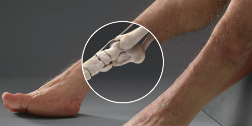
Here’s how nightmarish humans would look if our bodies were designed to survive car crashes
Article by Chris Weller, Tech Insider & Business Insider
If you’re ever in a car with Graham, then don’t bother telling him to buckle his seat belt. His body is already designed to withstand high-speed impacts.
Designed by a trauma surgeon, an artist, and a crash investigator, Graham is a hypothetical scenario come to life. Supported by Australia’s Transport Accident Commission, the project is meant to highlight how vulnerable humans are to injury.
Graham, however, is not.
Keep reading
The 12 Cranial Nerves and Starbucks

When you walk into Starbucks and you..
I. Smell the coffee aroma (olfactory)..
II. Read the order menu from about 20 feet away (optic) then you..
III. Pupils constrict as you look at items, such as muffins, closer (oculomotor)..
IV. You look up at salesperson then down at your money as you pay (trochlear)..
V. You clench your teeth and touch your face when they called your drink (trigeminal)..
VI. You look side-to-side to see if anyone else has ordered the same drink (abdsucens)..
VII. You smile because you realize this IS your drink (facial), then..
VIII. You hear someone say “You can sit here, we are leaving” (auditory). As you sit..
IX. You taste the sweet whipped cream on the top of your drink (glossopharyngeal)..
X. You say “Ahhhh this is good!” (vagus)..
XI. You look at the person next to you because they heard you and then you shrug your shoulders (spinal accessory)..
XII. When they look away you stick your tongue at them (hypoglossal)!


Bonus comic!
Yahoo! Einstein was right again! :D We now have our first detection of gravitational waves!
http://www.nytimes.com/2016/02/12/science/ligo-gravitational-waves-black-holes-einstein.html?_r=0
http://www.space.com/17661-theory-general-relativity.html


Scientists use lasers to control mouse brain switchboard
Ever wonder why it’s hard to focus after a bad night’s sleep? Using mice and flashes of light, scientists show that just a few nerve cells in the brain may control the switch between internal thoughts and external distractions. The study, partly funded by the National Institutes of Health, may be a breakthrough in understanding how a critical part of the brain, called the thalamic reticular nucleus (TRN), influences consciousness.
“Now we may have a handle on how this tiny part of the brain exerts tremendous control over our thoughts and perceptions,” said Michael Halassa, M.D., Ph.D., assistant professor at New York University’s Langone Medical Center and a lead investigator of the study. “These results may be a gateway into understanding the circuitry that underlies neuropsychiatric disorders.”
The TRN is a thin layer of nerve cells on the surface of the thalamus, a center located deep inside the brain that relays information from the body to the cerebral cortex. The cortex is the outer, multi-folded layer of the brain that controls numerous functions, including one’s thoughts, movements, language, emotions, memories, and visual perceptions. TRN cells are thought to act as switchboard operators that control the flow of information relayed from the thalamus to the cortex.
“The future of brain research is in studying circuits that are critical for brain health and these results may take us a step further,” said James Gnadt, Ph.D., program director at NIH’s National Institute Neurological Disorders and Stroke (NINDS), which helped fund the study. “Understanding brain circuits at the level of detail attained in this study is a goal of the President’s Brain Research through Advancing Innovative Neurotechnologies (BRAIN) Initiative.”
To study the circuits, the researchers identified TRN cells that send inhibitory signals to parts of the thalamus known to relay visual information to the cortex. Using a technique known as multi-electrode recordings, they showed that sleep and concentration affected these cells in opposite ways.
They fired often when the mice were asleep, especially during short bursts of simultaneous brain cell activity called sleep spindles. These activity bursts briefly widen electrical brain wave traces making them look like spindles, the straight spikes with rounded bottoms used to make yarn. In contrast, the cells fired infrequently when the mice were tasked with using visual cues to find food. The results suggested that these cells blocked visual information from reaching the cortex during sleep and allowed its transmission when the mice were awake and attentive.
For Dr. Halassa, a practicing psychiatrist who treats schizophrenia, these surprising results may provide fundamental insights into how the brain controls information transmission, a process that is disrupted in patients with neuropsychiatric disorders. Previous studies suggested that people who experienced more spindles while sleeping were less susceptible to being disturbed by outside noises. Moreover, people with schizophrenia and autism spectrum disorder may experience fewer spindles.
“Spindles may be peepholes into the mysteries of these disorders,” said Dr. Halassa.
To test this idea, the researchers used optogenetics, a technique that introduces light-sensitive molecules into nerve cells. This allowed them to precisely control the firing patterns of visual TRN cells with flashes of laser light. The experiments were performed in well-rested as well as sleep-deprived mice. Similar to what is seen in humans, sleep deprivation can disrupt the ability of mice to focus and block out external distractions.
Well-rested mice needed just a second or two to find the food whereas sleep-deprived mice took longer, suggesting that lack of sleep had detrimental effects on their ability to focus. When the researchers used flashes of laser light to inhibit the firing of optogenetically engineered visual TRN cells in sleep-deprived mice, the mice found the food faster. In contrast, if they used optogenetics to induce sleep-like firing patterns in well-rested mice, then the mice took longer to find food.
“It’s as if with a flick of a switch we could alter the mental states of the mice and either mimic or cure their drowsiness,” said Dr. Halassa.
In a parallel set of experiments the researchers found neighbors of the visual TRN cells had very different characteristics. These neighboring cells control the flow of information to the cortex from limbic brain regions, which are involved with memory formation, emotions and arousal. The cells fired very little during sleep and instead were active when the mice were awake. Dr. Halassa thinks that their firing pattern may be important for the strengthening of new memories that often occurs during sleep. Combined, the results suggest that the TRN is divided into sub-networks that oversee discrete mental states. The researchers think understanding the sub-networks is an initial step in thoroughly exploring the role of the TRN in brain disorders.

Both hemispheres of the brain process numbers
Researchers of the Jena University (Germany) and of the Jena University Hospital located an important region for the visual processing of numbers in the human brain and showed that it is active in both hemispheres. In the ’Journal of Neuroscience’ the scientists published high resolution magnetic resonance recordings of this region.
The human brain works with division of labour. Although our thinking organ excels in displaying amazing flexibility and plasticity, typically different areas of the brain take over different tasks. While words and language are mainly being processed in the left hemisphere, the right hemisphere is responsible for numerical reasoning. According to previous findings, this division of labour originates from the fact that the first steps in the processing of letters and numbers are also located individually in the different hemispheres. But this is not the case, at least not when it comes to the visual processing of numbers.
Neuroscientists of the Friedrich Schiller University Jena and of the Jena University Hospital discovered that the visual processing of numbers takes place in a so-called ‘visual number form area’ (NFA) - in fact in both hemispheres alike. The Jena scientists were the first to publish high resolution magnetic resonance recordings showing the activity in this region of the brain of healthy test persons. The area is normally difficult to get access to.
The 'blind spot’ in the brain
In their study Dr. Mareike Grotheer and Prof. Dr. Gyula Kovács from the Institute for Psychology of Jena University as well as Dr. Karl-Heinz Herrmann from the Department of Radiology (IDIR) of the Jena University Hospital presented subjects with numbers, letters and pictures of everyday objects. Meanwhile the participants’ brain activity was recorded using magnetic resonance imaging (MRI). Thus the researchers were able to clearly identify the region in which the visual processing of numbers takes place. The small area at the underside of the left and right temporal lobe reacted with increased activity at the presentation of numbers. Letters and other images but also false numbers lead to a significantly lower brain activity in this area.
Although the Jena team already knew from other scientists’ previous research where they had to look for the area, a lot of developmental work went into the newly published story. “This region has been a kind of blind spot in the human brain until now,” Mareike Grotheer says. And here is why: Hidden underneath the ear and the acoustic meatus, surrounded by bone and air, previous MRI scans showed a number of artefacts and thus obstructed detailed research.
For their study the Jena scientists used a high-performance 3 tesla MRI scanner of the Institute of Diagnostic and Interventional Radiology (IDIR) of the Jena University Hospital. They recorded three-dimensional images of the brain of the test subjects at an unusually high spatial resolution and hence with only very few artefacts. In addition these recordings were spatially smoothed whereby the remaining 'white noise’ could be removed. This approach will help other scientists to investigate a part of the brain that until now had been nearly inaccessible. “In this region not only numbers are being processed but also faces and objects,” Prof. Kovács states.
-
 mindkeeper liked this · 9 months ago
 -
 spirfoterne liked this · 1 year ago
 -
 aki-far liked this · 1 year ago
 -
 earthangelsworld reblogged this · 2 years ago
 -
earthangelsworld liked this · 2 years ago
-
 dig92-sex-74dif liked this · 2 years ago
 -
 k-ads liked this · 3 years ago
 -
 sirelsewhere reblogged this · 3 years ago
 -
 rayleests reblogged this · 3 years ago
 -
 sairahs-thoughts liked this · 4 years ago
 -
 stuffuff reblogged this · 4 years ago
 -
 alejandropinpon liked this · 4 years ago
 -
 myqueerdear reblogged this · 4 years ago
 -
 im-nike-1111 reblogged this · 4 years ago
 -
d0ct0r0 liked this · 4 years ago
