New Technique Captures The Activity Of An Entire Brain In A Snapshot
New technique captures the activity of an entire brain in a snapshot
When it comes to measuring brain activity, scientists have tools that can take a precise look at a small slice of the brain (less than one cubic millimeter), or a blurred look at a larger area. Now, researchers at The Rockefeller University have described a new technique that combines the best of both worlds—it captures a detailed snapshot of global activity in the mouse brain.

(Image caption: Sniff, sniff: This density map of the cerebral cortex of a mouse shows which neurons get activated when the animal explores a new environment. The lit up region at the center (white and yellow) represents neurons associated with the mouse’s whiskers)
“We wanted to develop a technique that would show you the level of activity at the precision of a single neuron, but at the scale of the whole brain,” says study author Nicolas Renier, a postdoctoral fellow in the lab of Marc Tessier-Lavigne, Carson Family Professor and head of the Laboratory of Brain Development and Repair, and president of Rockefeller University.
The new method, described in Cell, takes a picture of all the active neurons in the brain at a specific time. The mouse brain contains dozens of millions of neurons, and a typical image depicts the activity of approximately one million neurons, says Tessier-Lavigne. “The purpose of the technique is to accelerate our understanding of how the brain works.”
Making brains transparent
“Because of the nature of our technique, we cannot visualize live brain activity over time—we only see neurons that are active at the specific time we took the snapshot,” says Eliza Adams, a graduate student in Tessier-Lavigne’s lab and co-author of the study. “But what we gain in this trade-off is a comprehensive view of most neurons in the brain, and the ability to compare these active neuronal populations between snapshots in a robust and unbiased manner.”
Here’s how the tool works: The researchers expose a mouse to a situation that would provoke altered brain activity—such as taking an anti-psychotic drug, brushing whiskers against an object while exploring, and parenting a pup—then make the measurement after a pause. The pause is important, explains Renier, because the technique measures neuron activity indirectly, via the translation of neuronal genes into proteins, which takes about 30 minutes to occur.
The researchers then treat the brain to make it transparent—following an improved version of a protocol called iDISCO, developed by Zhuhao Wu, a postdoctoral associate in the Tessier-Lavigne lab—and visualize it using light-sheet microscopy, which takes the snapshot of all active neurons in 3-D.
To determine where an active neuron is located within the brain, Christoph Kirst, a fellow in Rockefeller’s Center for Studies in Physics and Biology, developed software to detect the active neurons and to automatically map the snapshot to a 3-D atlas of the mouse brain, generated by the Allen Brain Institute.
Although each snapshot of brain activity typically includes about one million active neurons, researchers can sift through that mass of data relatively quickly if they compare one snapshot to another snapshot, says Renier. By eliminating the neurons that are active in both images, researchers are left only those specific to each one, enabling them to home in on what is unique to each state.
Observing and testing how the brain works
The primary purpose of the tool, he adds, is to help researchers generate hypotheses about how the brain functions that then can be tested in other experiments. For instance, using their new techniques, the researchers, in collaboration with Catherine Dulac and other scientists at Harvard University, observed that when an adult mouse encounters a pup, a region of its brain known to be active during parenting—called the medial pre-optic nucleus, or MPO—lights up. But they also observed that, after the MPO area becomes activated, there is less activity in the cortical amygdala, an area that processes aversive responses, which they found to be directly connected to the MPO “parenting region.”
“Our hypothesis,” says Renier, “is that parenting neurons put the brake on activity in the fear region, which may suppress aversive responses the mice may have towards pups.” Indeed, mice that are being aggressive to pups tend to show more activity in the cortical amygdala.
To test this idea, the next step is to block the activity of this brain region to see if this reduces aggression in the mice, says Renier.
The technique also has broader implications than simply looking at what areas of the mouse brain are active in different situations, he adds. It could be used to map brain activity in response to any biological change, such as the spread of a drug or disease, or even to explore how the brain makes decisions. “You can use the same strategy to map anything you want in the mouse brain,” says Renier.
More Posts from Science-is-magical and Others
Here are 17 jaw-dropping photos of space that show us just how small we really are:
This photo of the moon and Earth taken from the International Space Station.

A dwarf galaxy, about 11 million light-years away from us.

Earth as seen from the moon in 1968.

A cluster of stars, 20,000 light-years away from Earth.

The first flower grown in the International Space Station, photographed by astronaut Scott Kelly.

Saturn, seen through an infared filter.

These visible “loops” on the surface of the sun can reach up to 15 times the diameter of Earth in height.

The Northen Lights just North of Chicago, viewed from the International Space Station.

The Quintuplet Cluster, located 100 light-years from the center of our galaxy.

Pluto and one of its moons, Charon.

The Great Pyramids of Giza, seen from space.

Astronaut Bruce McCandless maneuvering, untethered, above Earth in 1984.

Galaxy NGC 6240, 400 million light-years away from Earth.

Palomar 12, a cluster of stars on the outskirts of the Milky Way.

The remnants of an exploded star.

New York City, seen from the International Space Station.

And the remains of a supernova whose explosion may have been seen almost 2,000 years ago by Chinese astronomers.
Follow @the-future-now


Birds developed the unique vocal organ that enables them to sing more than 66 million years ago when dinosaurs walked the Earth, a new fossil discovery has shown.
But the earliest syrinx, an arrangement of vibrating cartilage rings at the base of the windpipe, was still a long way from producing the lilting notes of a song thrush or blackbird.
Scientists believe the extinct duck and goose relative that possessed the organ was only capable of making honking noises.
The bird, Vegavis iaai, lived during the Cretaceous era. Although its fossil bones were unearthed from Vega Island in Antarctica in 1992, it was not until three years ago that experts spotted the syrinx.
All birds living today are descended from a particular family of dinosaurs that developed feathers and the ability to fly.
The new discovery suggests the syrinx is another hallmark of birds that was absent from non-avian dinosaurs…


Machine Learning: An In-Depth, Non-Technical Guide
A good introductory text in five parts:
Overview, goals, learning types, and algorithms
Data selection, preparation, and modeling
Model evaluation, validation, complexity, and improvement
Model performance and error analysis
Unsupervised learning, related fields, and machine learning in practice
By Alex Castrounis - {InnoArchiTech}

Glutamate, an essential food for the brain
Glutamate is an amino acid with very different functions: in the pancreas, it modulates the activity of the pancreatic ß-cells responsible for insulin production, whereas in the brain it is the main excitatory neurotransmitter. In recent years, it has been suspected to play an additional role in the functioning of the brain. By discovering how the brain uses glutamate to produce energy, researchers at the University of Geneva (UNIGE) confirm this hypothesis and highlight unexpected links with the rest of the body. To read in Cell Reports.
Unlike other organs, the brain cannot draw its energy from lipids, an energy resource widely present in the body. The blood-brain barrier, which protects it from the pathogens and toxins circulating in the blood, indeed limits the passage of these lipids. Moreover, while most of the organs in the human body have the ability to store glucose by increasing their mass, the brain, prisoner of the cranial bones, cannot count on these variations in volume. Unable to store its food, it depends on sugar supplied in real-time by the rest of the body. This distribution of energy is controlled by the liver.
Pierre Maechler, professor at the Faculty of Medicine at UNIGE, and his team therefore decided to verify if glutamate was indeed an energy source for the brain. To do so, the researchers analyzed the role of the glutamate dehydrogenase enzyme in the brain. In mutant form, this enzyme, encoded by the Glud1 gene, is responsible for a congenital hyperinsulinism syndrome, a severe disease affecting at the same time the endocrine pancreas, the liver and the brain. Individuals affected by this syndrome suffer from intellectual disability and have a high risk of epilepsy. “We have suppressed the Glud1 gene in the brain of mice. In the absence of glutamate dehydrogenase, we observed that the brain was no longer able to convert glutamate into energy, even though the amino acid was present in the brain,” explains Melis Karaca, first author of this study.
Priority to the brain
Devoid of the energy supplied by cerebral glutamate, the brain sends signals to the liver to requisition a compensatory proportion of glucose, at the expense of the rest of the body. This is why the transgenic mice also showed a growth deficit and muscle atrophy. “This clearly shows how the brain works in a just-in-time manner and that each percent of energy resources is essential for its proper functioning,” highlights Professor Pierre Maechler. “If a part of this energy disappears, the brain serves itself first and the rest of the body suffers. The liver must then make more glucose by drawing upon muscle protein, resulting in loss of muscle mass. Knowing that the brain uses glutamate as an energy resource allows us to reflect on other ways to overcome a potential shortfall. ”
Scientists also suspect a correlation between the Glud1 gene and some neurodevelopmental disorders, particularly epilepsy and schizophrenia. They are currently pursuing their research by introducing in mice the same Glud1 mutation detected in epileptic patients. At the same time, another group is working with schizophrenic patients to assess the way their brain uses glutamate.
A puzzling expected value
Pick a (uniformly) random real number from the unit interval [0,1] and repeat this until the sum of all chosen numbers exceeds 1. What is the expected number of real values you will pick?
The quite surprising answer is Eulers constant, e ≈ 2.71828.
A demonstration can be found on Wolfram MathWorld.


Better late than never!
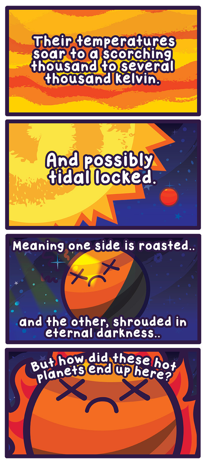
This week’s entry: Hot Jupiters
http://www.space.com/32011-extremely-hot-and-fast-planets-seem-to-defy-logic.html
https://astrobites.org/2015/03/04/hot-jupiters-are-very-bad-neighbors/
-
 science-is-magical reblogged this · 8 years ago
 -
 facesofconsciousness reblogged this · 8 years ago
 -
 sienfilos reblogged this · 8 years ago
 -
 kingdomofrosesqueenidun reblogged this · 8 years ago
 -
 carldepolairbeer-blog liked this · 8 years ago
 -
 rowrrr liked this · 8 years ago
 -
 ibiofeedback reblogged this · 8 years ago
 -
 annamariexx32921 liked this · 8 years ago
 -
 the-faktory liked this · 8 years ago
 -
 1grammiefan liked this · 8 years ago
 -
 mollyfamous reblogged this · 8 years ago
 -
 taraneecooking reblogged this · 8 years ago
 -
 modernstoneage liked this · 8 years ago
 -
 pbarrr liked this · 8 years ago
 -
 brittaniahart liked this · 8 years ago
 -
 mlle-rousse liked this · 8 years ago
 -
 nikkidetko reblogged this · 8 years ago
 -
ronin9gin liked this · 8 years ago
-
 pokecatt liked this · 8 years ago
 -
 ihazcatmunster liked this · 8 years ago
 -
 nurnielfa liked this · 8 years ago
 -
 queenofeire liked this · 8 years ago
 -
coolasp liked this · 8 years ago
-
 burntgazpacho reblogged this · 8 years ago
 -
 icarus-never-burned reblogged this · 8 years ago
 -
 disco-dancer-donna liked this · 8 years ago
 -
 hasbro-necromancer liked this · 8 years ago
 -
 leftfootism liked this · 8 years ago
 -
 thepensivenotebook-blog reblogged this · 8 years ago
 -
 welovesome liked this · 8 years ago
 -
 virtual-particle liked this · 8 years ago
 -
 jsprags liked this · 8 years ago
 -
 adorable-amygdala liked this · 8 years ago
