It’s Time For #TrilobiteTuesday! During Their Lengthy Trek Through Time, Trilobites Existed In An Almost

It’s time for #TrilobiteTuesday! During their lengthy trek through time, trilobites existed in an almost dizzying array of sizes and shapes. Perhaps no other creature in the entire history of the earth has ever displayed the diversity of design shown by these singularly distinctive arthropods. But at their heart (and yes, trilobites apparently did possess primitive but effective cardio-respiratory systems), they were all remarkably similar. Named not, as is generally surmised, for their three main body segments – cephalon (head), thorax (body) and pygidium (tail) – but rather for the three lobes that longitudinally divided their dorsal exoskeleton. Whether they were Cambrian Olenellids – such as this Olenellus romensis from Alabama – or Devonian Phacopids, most trilobites presented a fundamentally analogous body design. Such characteristics as occipital lobes, anterior margins and facial sutures (which allowed early trilobites to shed their molting shell), were shared by the majority of trilobite species, as were such exotic-sounding features as axial rings, articulating facets and pleural spines.
More Posts from Contradictiontonature and Others


Fungal tissues – the fungal mantle around the root tip and the fungal network of tendrils that penetrates the root of plants, or Hartig Net, between Pinus sylvestris plant root cells – in green. Ectomycorrhizal (ECM) fungi help trees tolerate drought and boost the productivity of bioenergy feedstock trees, including poplar and willow.
Via Berkeley Lab: The sclerotia are in the soil!
More: How Fungi Help Trees Tolerate Drought (Joint Genome Institute)
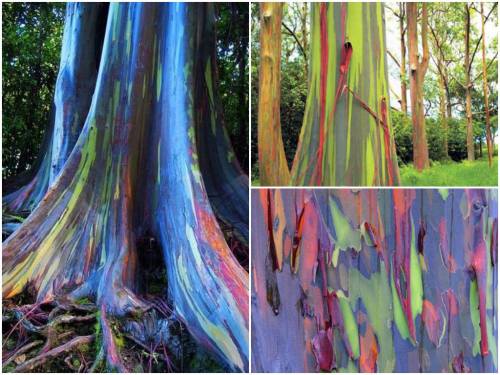
These are rainbow eucalyptus trees (Eucalyptus deglupta) and hail from the Philippine Islands.
The trees get their name from the striking colours observed on their trunks and limbs. Although it may look like someone took a paintbrush to them, these colours are entirely natural. Unlike most trees, the rainbow eucalyptus does not have a thick, cork-like layer of bark on its trunk. The bark is smooth and as it grows it ‘exfoliates’ layers of spent tissue. This exfoliation technique occurs at different stages and in different zones of the tree.
Keep reading

(Image caption: An fMRI scan shows regions of the brain that become active when devoutly religious study participants have a spiritual experience, including a reward center in the brain, the nucleus accumbens. Credit: Jeffrey Anderson)
This is your brain on God
Religious and spiritual experiences activate the brain reward circuits in much the same way as love, sex, gambling, drugs and music, report researchers at the University of Utah School of Medicine. The findings were published in the journal Social Neuroscience.
“We’re just beginning to understand how the brain participates in experiences that believers interpret as spiritual, divine or transcendent,” says senior author and neuroradiologist Jeff Anderson. “In the last few years, brain imaging technologies have matured in ways that are letting us approach questions that have been around for millennia.”
Specifically, the investigators set out to determine which brain networks are involved in representing spiritual feelings in one group, devout Mormons, by creating an environment that triggered participants to “feel the Spirit.” Identifying this feeling of peace and closeness with God in oneself and others is a critically important part of Mormons’ lives — they make decisions based on these feelings; treat them as confirmation of doctrinal principles; and view them as a primary means of communication with the divine.
During fMRI scans, 19 young-adult church members — including seven females and 12 males — performed four tasks in response to content meant to evoke spiritual feelings. The hour-long exam included six minutes of rest; six minutes of audiovisual control (a video detailing their church’s membership statistics); eight minutes of quotations by Mormon and world religious leaders; eight minutes of reading familiar passages from the Book of Mormon; 12 minutes of audiovisual stimuli (church-produced video of family and Biblical scenes, and other religiously evocative content); and another eight minutes of quotations.
During the initial quotations portion of the exam, participants — each a former full-time missionary — were shown a series of quotes, each followed by the question “Are you feeling the spirit?” Participants responded with answers ranging from “not feeling” to “very strongly feeling.”
Researchers collected detailed assessments of the feelings of participants, who, almost universally, reported experiencing the kinds of feelings typical of an intense worship service. They described feelings of peace and physical sensations of warmth. Many were in tears by the end of the scan. In one experiment, participants pushed a button when they felt a peak spiritual feeling while watching church-produced stimuli.
“When our study participants were instructed to think about a savior, about being with their families for eternity, about their heavenly rewards, their brains and bodies physically responded,” says lead author Michael Ferguson, who carried out the study as a bioengineering graduate student at the University of Utah.
Based on fMRI scans, the researchers found that powerful spiritual feelings were reproducibly associated with activation in the nucleus accumbens, a critical brain region for processing reward. Peak activity occurred about 1-3 seconds before participants pushed the button and was replicated in each of the four tasks. As participants were experiencing peak feelings, their hearts beat faster and their breathing deepened.
In addition to the brain’s reward circuits, the researchers found that spiritual feelings were associated with the medial prefrontal cortex, which is a complex brain region that is activated by tasks involving valuation, judgment and moral reasoning. Spiritual feelings also activated brain regions associated with focused attention.
“Religious experience is perhaps the most influential part of how people make decisions that affect all of us, for good and for ill. Understanding what happens in the brain to contribute to those decisions is really important,” says Anderson, noting that we don’t yet know if believers of other religions would respond the same way. Work by others suggests that the brain responds quite differently to meditative and contemplative practices characteristic of some eastern religions, but so far little is known about the neuroscience of western spiritual practices.
The study is the first initiative of the Religious Brain Project, launched by a group of University of Utah researchers in 2014, which aims to understand how the brain operates in people with deep spiritual and religious beliefs.


This massive virus with its own immune system could hold the future of medicine
French researchers think they’ve found a giant virus big enough to house its own virus-killing devices using a system like CRISPR, and it could be a completely new form of life.
Called a mimivirus, it was first found growing in amoebae in a water tower. At four times the size of a typical virus, you can even see it under a light microscope
When the mimivirus encounters another virus, it stores some of the invader’s genetic material. That way, when it encounters the same kind of virus again, the MIMIVIRE system goes into gene-editing berserker mode, finding the key genes of the virus and cutting them to inert oblivion. This could have major applications.
Follow @the-future-now

Quote from Sau Lan Wu, scientist who discovered the #higgsboson. More quotes like this one to inspire you in my I Love Science Journal coming to stores in March. Preorder now on Amazon! #ilovescience #womeninscience #scientificliteracy

New insights into the molecular basis of memory
Scientists from the German Center for Neurodegenerative Diseases (DZNE) in Göttingen and Munich have shed new light on the molecular basis of memory. Their study confirms that the formation of memories is accompanied by an altered activity of specific genes. In addition, they found an unprecedented amount of evidence that supports the hypothesis that chemical labels on the backbone of the DNA (so-called DNA methylation) may be the molecular basis of long-term memory. These findings are reported in “Nature Neuroscience”.
The brain still harbours many unknowns. Basically, it is assumed that it stores experiences by altering the connections between brain cells. This ability to adapt – which is also called “plasticity” – provides the basis for memory and learning, which is the ability to draw conclusions from memories. On a molecular scale these changes are mediated by modifications of expression of specific genes that as required strengthen or weaken the connections between the brain cells.
In the current study, a research team led by Dr. Stefan Bonn and Prof. André Fischer from Göttingen, joined forces with colleagues from the DZNE’s Munich site, to examine how the activity of such genes is regulated. The scientists stimulated long-term memory in mice, by training the animals to recognise a specific test environment. Based on tissue samples, the researchers were able to discern to what extent this learning task triggered changes in the activity of the genes in the mice’s brain cells. Their focus was directed on so-called epigenetic modifications. These modifications involve the DNA and DNA associated proteins.
Epigenetic modifications
“The cell makes use of various mechanisms in order to turn genes on or off, without altering the DNA sequence itself. It’s called ‘epigenetics’,” explains Dr. Magali Hennion, a staff member of the research group of Stefan Bonn.
In principle, gene regulation can happen through methylation, whereby the backbone of the DNA is chemically labeled at specific sites. Changes in the proteins called histones that are packaging the DNA may also occur.
Hennion: “Research on epigenetic changes that are related to memory processes is still at an early stage. We look at such features, not only for the purpose of a better understanding of how memory works. We also look for potential targets for drugs that may counteract memory decline. Ultimately, our research is about therapies against Alzheimer’s and similar brain diseases.“
A code for memory contents?
In the current study the researchers found modifications, both of the histones as well as of the methylation of the DNA. However, histone modifications had little effect on the activity of genes involved in neuroplasticity. Furthermore, Bonn and his colleagues not only discovered epigenetic modifications in nerve cells, but also in non-neuronal cells of the brain.
“The relevance of non-neuronal cells for memory, is an interesting topic that we will continue to pursue“, says André Fischer, site speaker for the DZNE in Göttingen and professor at the University Medical Center Göttingen (UMG). “Furthermore, our observations suggest that neuroplasticity is to a large extent regulated by DNA methylation. Although this is not a new hypothesis, our study provides an unprecedented amount of supporting evidence for this. Thus, methylation may indeed be an important molecular constituent of long-term memory. In such a case, methylation could be a sort of code for memory content and a potential target for therapies against Alzheimer’s disease. This is an aspect that we specifically want to focus on, in further studies.”

Quote by #rosalindfranklin How do you make science a part of your life? What are you doing to fight for scientific literacy? More quotes and questions in my #ilovescience journal. #womeninscience #scientificliteracy

Poison in the Brain: Toxic Structures in Neuronal Nuclei of Alzheimer’s Patients
Spherical structures in the nucleus of nerve cells, so-called nuclear spheres, are suspected to trigger Alzheimer’s disease. A team headed by Dr Thorsten Müller from the research group Cell Signaling in Neurodegeneration has for the very first time demonstrated the presence of the presumably toxic protein aggregates in the human brain. The researchers from Ruhr-Universität Bochum have published their article in the journal Neurobiology of Aging.
The team compared brain samples from Alzheimer’s patients with those of the healthy individuals in the same age group. The result: in the samples taken from Alzheimer’s patients, the number of nuclear spheres was much higher than in those taken from healthy study participants.
Moreover, the group from Bochum analysed in what way nuclear spheres are generated. It was demonstrated in experiments with cell cultures that the amyloid precursor protein (APP) plays a crucial role in this process. APP has long been associated with Alzheimer’s disease. The researchers observed that nuclear sphere generation preferably takes place, if the amyloid precursor protein carries no phosphate group in a specific amino acid. An APP cleavage product, moreover, is contained in the nuclear spheres.
“Extensive nuclear sphere generation in the human Alzheimer’s brain” by Katharina Kolbe, Hassan Bukhari, Christina Loosse, Gregor Leonhardt, Annika Glotzbach, Magdalena Pawlas, Katharina Hess, Carsten Theiss, and Thorsten Müller in Neurobiology of Aging. Published online August 18 2016 doi:10.1016/j.neurobiolaging.2016.08.016
-
 metalzoic liked this · 6 years ago
 -
 eykonto reblogged this · 6 years ago
 -
eykonto liked this · 6 years ago
-
 i-was-supposed-to-have-a-twin liked this · 6 years ago
 -
 bolaneno liked this · 6 years ago
 -
 fancysaurus liked this · 8 years ago
 -
 paleontologylife reblogged this · 8 years ago
 -
paleontologylife liked this · 8 years ago
-
 fkinghippi liked this · 8 years ago
 -
 philosophical-fantastical liked this · 8 years ago
 -
 miguelangel1974 liked this · 8 years ago
 -
 yougotgeminilaser reblogged this · 8 years ago
 -
 delicateeaglepaper liked this · 8 years ago
 -
 wemblingfool reblogged this · 8 years ago
 -
 memeologist-who-tries liked this · 8 years ago
 -
 thanatoast250 reblogged this · 8 years ago
 -
 pikuotaku liked this · 8 years ago
 -
 sadfacez liked this · 8 years ago
 -
 sibuyanensis liked this · 8 years ago
 -
 prettypangolins reblogged this · 8 years ago
 -
 hopelesswanderer-danilion liked this · 8 years ago
 -
 hslmoreno liked this · 8 years ago
 -
 phylosopherstoned liked this · 8 years ago
 -
 cool-david-stafford liked this · 8 years ago
 -
 itsrebfergworld liked this · 8 years ago
 -
 jbail86 liked this · 8 years ago
 -
 treyfla liked this · 8 years ago
 -
 roseh23 liked this · 8 years ago
 -
 kurtmn liked this · 8 years ago
 -
 shatovthings liked this · 8 years ago
 -
 cowskier99 liked this · 8 years ago
 -
 mgeverroad-blog liked this · 8 years ago
 -
 robtheprog-blog liked this · 8 years ago
 -
 bibliophiliosaurus reblogged this · 8 years ago
 -
 clever-boxing liked this · 8 years ago
 -
texaslovinmimi liked this · 8 years ago
-
 nina-brujita reblogged this · 8 years ago
 -
nina-brujita liked this · 8 years ago
-
 biologynerd reblogged this · 8 years ago
 -
 formulafun liked this · 8 years ago
 -
 aintnobodysbusinessifido liked this · 8 years ago
 -
 amphetamine--logic-blog liked this · 8 years ago
 -
 andriacrowjoy liked this · 8 years ago
 -
 annilove8 liked this · 8 years ago
A pharmacist and a little science sideblog. "Knowledge belongs to humanity, and is the torch which illuminates the world." - Louis Pasteur
215 posts