Today Is World Anaesthesia Day! Here’s A Look At The Chemistry Behind A Range Of Anaesthetics. More


Today is World Anaesthesia Day! Here’s a look at the chemistry behind a range of anaesthetics. More info here and here.
More Posts from Contradictiontonature and Others

After the news of an accident in a French drug trial on Friday, you might be wondering what drug trials entail. Here’s a summary sheet on the drug discovery process to clear things up! http://wp.me/p4aPLT-1EZ
Sounds, such as music and noise, are capable of reliably affecting individuals’ moods and emotions, possibly by regulating brain dopamine, a neurotransmitter strongly involved in emotional behavior and mood regulation.


Image of the Week – February 13, 2017
CIL:40984 - http://www.cellimagelibrary.org/images/40984
Description: Montage image of a brain stem from a Brainbow transgenic mouse. In Brainbow mice, neurons randomly choose combinations of red, yellow and cyan fluorescent proteins, so that they each glow a particular color. This provides a way to distinguish neighboring neurons and visualize brain circuits. These are large caliber axons of the auditory pathway. First Prize, 2007 Olympus BioScapes Digital Imaging Competition. For additional details see: Livet J, Weissman TA, Kang H, Draft RW, Lu J, Bennis RA, Sanes JR, Lichtman JW. Nature. 2007 Nov 1;450(7166):56-62.
Authors: Jean Livet and the 2007 Olympus BioScapes Digital Imaging Competition®
Licensing: Attribution Non-Commercial No Derivatives: This image is licensed under a Creative Commons Attribution, Non-Commercial, No Derivatives License

Even after someone is declared dead, life continues in the body, suggests a surprising new study with important implications.
Gene expression — when information stored in DNA is converted into instructions for making proteinsor other molecules — actually increases in some cases after death, according to the new paper, which tracked postmortem activity and is published in the journal Open Biology.
“Not all cells are ‘dead’ when an organism dies,” senior author Peter Noble of the University of Washington and Alabama State University told Seeker. “Different cell types have different life spans, generation times and resilience to extreme stress.”
In fact, some cells seem to fight to live after the organism has died.
“It is likely that some cells remain alive and are attempting to repair themselves, specifically stem cells,” Noble said.


Solidification of liquid Gallium
Gallium is a chemical element with symbol Ga and atomic number 31. Gallium is a soft, silvery metal, and elemental gallium is a brittle solid at low temperatures, and melts at 29.76 °C (85.57 °F) (slightly above room temperature). Elemental gallium is not found in nature, but it is easily obtained by smelting.
Gallium metal expands by 3.1% when it solidifies, and therefore storage in either glass or metal containers are avoided, due to the possibility of container rupture with freezing. Gallium shares the higher-density liquid state with only a few materials, like water, silicon,germanium, bismuth, and plutonium.
Giffed by: rudescience From: This video
Thumpety thump thump thumpety thump thump look at Kinesin go
Myosin, kinesin, and dynein are important proteins governing internal transport. Myosin attached to organelles associates with actin microfilaments to enable the continuous flow of cytoplasm called cytoplasmic streaming.
Kinesins and dynein enable the movement of organelles along microtubules. They attach and move along microtubules. Most kinesins transport organelles from the center towards the periphery of the cell, anterograde transport. Dynein, and a few types of kinesins transport towards the cell center, retrograde transport.
(via thelifeofapremed)

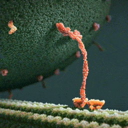
Scientists show how drug binds with ‘hidden pocket’ on flu virus
A new study led by scientists at The Scripps Research Institute (TSRI) is the first to show exactly how the drug Arbidol stops influenza infections. The research reveals that Arbidol stops the virus from entering host cells by binding within a recessed pocket on the virus.
The researchers believe this new structural insight could guide the development of future broad-spectrum therapeutics that would be even more potent against influenza virus.
“This is a very interesting molecule, and now we know where it binds and precisely how it works,” said study senior author Ian Wilson, Hanson Professor of Structural Biology, chair of the Department of Integrative Structural and Computational Biology and member of the Skaggs Institute for Chemical Biology at TSRI.
The study was published in the journal Proceedings of the National Academy of Sciences.
Rameshwar U. Kadam, Ian A. Wilson. Structural basis of influenza virus fusion inhibition by the antiviral drug Arbidol. Proceedings of the National Academy of Sciences, 2016; 201617020 DOI: 10.1073/pnas.1617020114
This is a 3-dimensional illustration showing the different features of an influenza virus, including the surface proteins hemagglutinin (HA) and neuraminidase (NA)/CDC

(Image caption: The synapses of pyramid cells in the cerebral cortex form functional groups. Some of the related synapses are shown in green in the reconstruction. Credit: © MPI of Neurobiology / Scheuss)
Neurons form synapse clusters
The cerebral cortex resembles a vast switchboard. Countless lines carrying information about the environment, for example from the sensory organs, converge in the cerebral cortex. In order to direct the flow of data into meaningful pathways, the individual pyramidal cells of the cerebral cortex act like miniature switchboard operators. Each cell receives information from several thousand lines. If the signals make sense, the line is opened, and the information is relayed onward. Scientists at the Max Planck Institute of Neurobiology in Martinsried have now shown for the first time that contact points between specific neuron types are clustered in groups on the target neuron. It is probable that signals are coordinated with each other in this way to make them more “convincing”.
The cells of the cerebral cortex have a lot to do. They process various types of information depending on the area in which they are located. For example, signals from the retina arrive in the visual cortex, where, among other things, the motion of objects is detected. The pyramidal cells of the cerebral cortex receive information from other cells through thousands of contact points called synapses. Depending on where, how many and how often synapses are activated, the cell relays the signal onward – or not.
Information is passed on in the form of electrical signals. The neurobiologists were able to measure these signals at various contact points of the neuron. “The exciting thing is that the signals that a cell receives from, say, ten simultaneously active synapses can be greater than the sum of the signals from the ten individual synapses,” says Volker Scheuss, summarizing the basis of his recently published study. “However, until now it was unclear whether this phenomenon can be explained by a specific arrangement of synapses on pyramidal cells.”
By combining modern methods, the neurobiologists in Tobias Bonhoeffer’s Department have analysed the arrangement of synapses. They were able to selectively activate a specific type of pyramid cell in brain slices from mice using optogenetics. Thanks to simultaneous “calcium imaging”, they were then able to observe and record the activity of individual synapses under a two-photon microscope. In this way, they succeeded in showing for the first time how synapses are arranged with respect to each other.
The result of such synapse mapping analysed with a newly developed algorithm was clear: The synapses of pyramidal cells form clusters consisting of 4 to 14 synapses arranged within an area of less than 30 micrometres along the dendrite. “The existence of these clusters suggests that the synapses interact with each other to control the strength of the combined signal,” explains Onur Gökçe, author of the study. This is the first anatomical explanation for the disproportionate strength of clustered synapse signals in comparison to the individual signals – a finding known from activity measurements. The observation in layer 5 pyramidal cells was of particular interest, as the activity of these cells oscillates synchronously. “This rhythmic activity, which probably influences the processing of visual information, could synchronously activate synapse clusters, thus boosting the overall signal received,” says Scheuss.
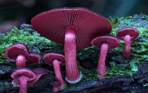
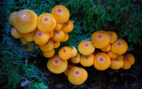
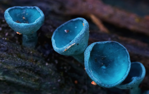
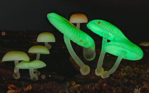

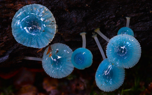
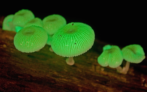
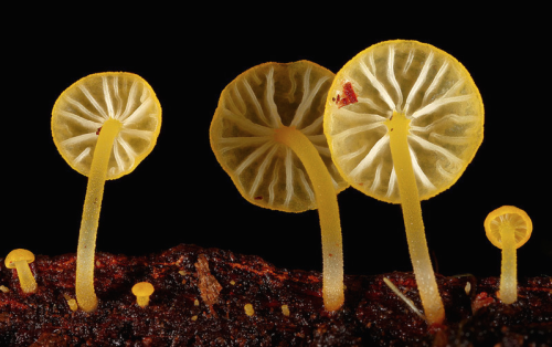
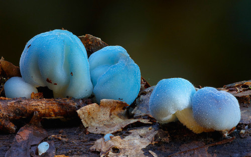
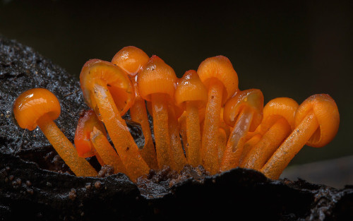
a mushroom rainbow to put the fun in fungi. cause they don’t need psilocybin to be magic. and though some mushrooms are coloured as a toxicity warning to predators, many others are brightly coloured to instead attract potential spore dispersers. see this for more on the bioluminescent mushrooms seen here. (photos)

IN THE MIX
Matti Koivisto, an undergraduate student working in the laboratory of Kari Haajanen at Turku University of Applied Sciences, designs solutions similar to this one to detect the bacteria Escherichia coli. He uses a dye called rhodamine 6G, which has a strong orange color when dissolved in ethanol. In solution, the dye separates into negatively and positively charged ions, the latter of which are largely responsible for the dye’s color. Negatively charged molecules on the outer membranes of E. coli attract the dye’s positive ions. This interaction causes the dye to change color to a pinkish hue, and the level of color change allows Koivisto to gauge how many of the bacteria are present.
Submitted by Matti Koivisto
Do science. Take photos. Make money: Enter our monthly photo contest here for your chance to win!
Related C&EN content:
How dyes in mouse feces help track insects
Using an ink-jet printer to simplify analytical chemistry
-
 thiawen liked this · 7 months ago
 -
 demise-of-soul liked this · 7 months ago
 -
 atomicstarburstlabware reblogged this · 7 months ago
 -
 dragoodanimala liked this · 4 years ago
 -
 defendersofshield liked this · 5 years ago
 -
 inboundflight liked this · 5 years ago
 -
 the-big-bad-litle-wolf liked this · 5 years ago
 -
 aestheticbit-blog liked this · 6 years ago
 -
 grungy-blue-hipster liked this · 6 years ago
 -
 m52hspringapple liked this · 6 years ago
 -
 itsboomdizzle reblogged this · 6 years ago
 -
fractal-fluctuation liked this · 6 years ago
-
 thievesandtraitors liked this · 7 years ago
 -
 izy-i-blog liked this · 7 years ago
 -
 king-flippy liked this · 7 years ago
 -
 sydneyruthwood reblogged this · 7 years ago
 -
sydneyruthwood liked this · 7 years ago
-
 kimiishi liked this · 7 years ago
 -
 tiniestrhino liked this · 7 years ago
 -
 behavioralgarbage reblogged this · 7 years ago
 -
 alchemystik reblogged this · 7 years ago
 -
 katyest liked this · 7 years ago
 -
 alligator-alley-blog reblogged this · 7 years ago
 -
 davidgandytt liked this · 7 years ago
 -
 steffi1986 liked this · 7 years ago
 -
 poshdetective liked this · 7 years ago
 -
 hollygopossumlovesj2 liked this · 7 years ago
 -
 soysaucevictim reblogged this · 7 years ago
 -
vetreferences reblogged this · 7 years ago
-
 plovertime liked this · 7 years ago
 -
 sparrowhawkandco reblogged this · 7 years ago
 -
 devonoff reblogged this · 7 years ago
 -
 joeygrump reblogged this · 7 years ago
 -
 ganbaregirl reblogged this · 7 years ago
 -
 hazel-sage liked this · 7 years ago
 -
anangryplatypus-blog liked this · 8 years ago
-
 bluebabadee-blog liked this · 8 years ago
 -
 studywithfox reblogged this · 8 years ago
 -
 champawattigress liked this · 8 years ago
 -
 drewsfedora reblogged this · 8 years ago
A pharmacist and a little science sideblog. "Knowledge belongs to humanity, and is the torch which illuminates the world." - Louis Pasteur
215 posts