What Is Brightness?

What is Brightness?
There has always been this nudge,right. The nudge to know what IS brightness. The subtle divergence between what we perceive as dim and bright. What it really means!
The brilliance of the sun whose light emerges afar blinds us but yet the quotidian florescent light seems to oblivious to us. Why this madness ! It doesn’t make sense to me.
Light as a particle

Two light bulbs 100W and 20W respectively. It is obvious that the 100 W glows brighter than its counterpart.
This is so because each photon ( a particle ) carries with it an amount of energy proportional to its frequency; E=hν. The energy dissipated per unit area is the energy per photon times the photons per unit area per second.
The 100 W bulb emits more photons per second than the 20 W bulb.

In this model, the photoreceptors in your eye undergo chemical reactions as a result of absorbing photons. The more photons absorbed per second, the brighter the light appears.
Light as a wave

In the picture of light as an electromagnetic wave, the energy carried by the light is proportional to the square of the wave’s amplitude.
In this model, the photo-receptors in your eye are oscillators. What is oscillating? Electric charge.
Charges are accelerated in response to the electric field of the light: the greater the electric field (or amplitude), the greater the amplitude of the oscillation, and the greater the electric currents in your eye (and the greater the brightness).
The human eye is truly a marvel. The level of serenity it brings to life is just enthralling.Have a great day!
- Post adapted from this stackexchange.
More Posts from Theperpetualscholar and Others

Time flies when you’re diverging at approximately 2% per million years
Anyway if you’re a nerd check out the coolest website ever: http://www.timetree.org/
You can input things like “dog” and “oyster mushroom” and it’ll tell you how long ago they diverged based on current data averages.
Transcript below the cut.
Patreon | Instagram | Facebook | Twitter | deviantArt
Keep reading


Climate Change


It’s #InternationalWomensDay! Here are twelve pioneering female chemists. Larger image & downloadable poster: http://wp.me/p4aPLT-2ra
New Evidence in Mice That Cocaine Makes Brain Cells Cannibalize Themselves
Working with mice, researchers at Johns Hopkins have contributed significant new evidence to support the idea that high doses of cocaine kill brain cells by triggering overactive autophagy, a process in which cells literally digest their own insides. Their results, moreover, bring with them a possible antidote, an experimental compound dubbed CGP3466B.
A summary of the study, which also found signs of autophagy in the brain cells of mice whose mothers received cocaine while pregnant, was published online the week of Jan. 18 in the Proceedings of the National Academy of Sciences.

(Image caption: A neural cell from a mouse brain shows much larger, more numerous vacuoles (orange) after 3 hours of treatment with cocaine than untreated cells. Credit: Prasun Guha, Maged Harraz, Solomon Snyder)

(Image caption: An untreated neural cell from a mouse brain shows no vacuoles. Credit: Prasun Guha, Maged Harraz, Solomon Snyder)
“We performed ‘autopsies’ to find out how cells die from high doses of cocaine,” says Solomon Snyder, M.D., professor of neuroscience at the Johns Hopkins University School of Medicine. “That information gave us immediate insight into how we might use a known compound to interfere with that process and prevent the damage.”
After discovering in 1990 that brain cells use the gas nitric oxide to communicate, Snyder and his research team have spent decades studying its impact. In 2013, the team found that nitric oxide is involved in cocaine-induced cell death through its interactions with GAPDH, an enzyme, but didn’t learn how precisely the cells were dying.
To find out, the research team examined nerve cells from mouse brains for clues. Snyder says cells, like whole animals, can die from extreme temperatures, toxins and physical trauma, but can also commit “suicide” in three ways that are chemically programmed and controlled by different proteins.
One such way is autophagy, a normal and much-needed cellular “cleanup process” that rids cells of debris that accumulates in membrane-enclosed vacuoles, or “bags” within the cell. These bags fuse with other bags, enzyme-rich lysosomes, which are filled with acids that degrade the contents of the vacuoles. Only when this process accelerates and spins out of control does it cause cell death, Snyder explains.
By measuring changes in the levels of proteins that control each cell death program and by observing the cells’ physical changes, the team saw clearly that cocaine causes neuronal cell death through out-of-control autophagy. That confirmed previous results from two other groups that found cocaine-induced autophagy in astrocytes and microglia, which are neuron support cells.
“A cell is like a household that is constantly generating trash,” says Prasun Guha, Ph.D., postdoctoral fellow at Johns Hopkins and lead author of the paper. “Autophagy is the housekeeper that takes out the trash — it’s usually a good thing. But cocaine makes the housekeeper throw away really important things, like mitochondria, which produce energy for the cell.”
Because the team already knew that nitric oxide and GAPDH were involved in the process, they tested the ability of the compound CGP3466B, known to disrupt nitric oxide/GAPDH interactions, to halt cocaine-induced autophagy. They also tested other chemicals known to prevent the other two forms of cellular suicide, but only CGP3466B protected mouse nerve cells in the brain from death by cocaine.
According to previous research from the same team, CGP3466B was also able to rescue the brain cells of live mice from the deadly effects of cocaine, but they had not connected the phenomenon to autophagy. When the scientists recently gave mice a single dose of cocaine and looked for signs of autophagy in their brain cells, they detected autophagy-associated proteins and changes in vacuoles in adults and in mouse pups whose mothers had received cocaine while pregnant.
“Since cocaine works exclusively to modulate autophagy versus other cell death programs, there’s a better chance that we can develop new targeted therapeutics to suppress its toxicity,” says Maged M. Harraz, Ph.D., a research associate at Johns Hopkins and lead co-author of the paper.
Snyder says the team hopes its work will eventually lead to treatments that protect adults and infants from the devastating effects of cocaine on the brain. Since CGP3466B has already been tested in phase II clinical trials to (unsuccessfully) treat Parkinson’s disease and ALS, it is known to be safe for humans, but the researchers caution that many more years of studies are needed to definitively show whether it is effective for preventing cocaine damage, first in mice, then in humans. They also want to create and test derivatives of CGP3466B to learn more about cocaine-induced autophagy and see if cocaine is killing any cells outside the brain.

In the left column starting at the top they are Orchard Oriole, Red-winged Blackbird, Eastern Towhee, Northern Cardinal, American Robin and Gray Catbird. And in the right column from the top they are Blue Jay, Carolina Chickadee, Song Sparrow, Field Sparrow, Chipping Sparrow and Seaside Sparrow.
source:

The study of life. Biology in Daily Life. 1953.

Hello everyone.
Apologies for the long/unexplained absence. I’ve not been doing very well recently and I haven’t quite had the time to be posting. However, a small update is that I’m struggling very much with my mental health but my studies are going very well. I just wanted to thank you all for your kindness and support and I will try update more regularly from now on!
I hope you are all well and looking after yourself, and I’m sending love, hugs and good study vibes 💗
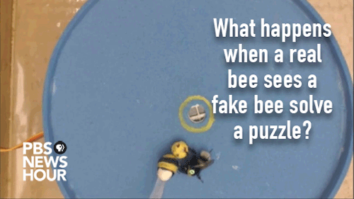
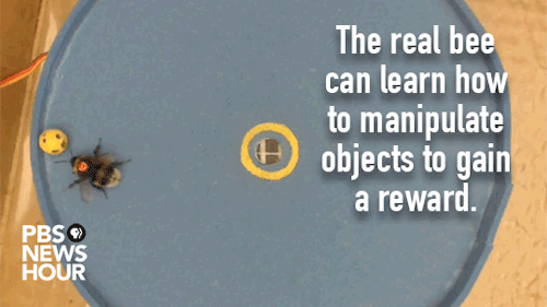
What does it take to teach a bee to use tools? A little time, a good teacher and an enticing incentive. Read more here: http://to.pbs.org/2mpRUAz
Credit: O.J. Loukola et al., Science (2017)
-
 jaibhagwan reblogged this · 4 years ago
 -
 go-wind-stuff reblogged this · 5 years ago
 -
 ginnysgotagun liked this · 6 years ago
 -
 ineverland reblogged this · 7 years ago
 -
 fables-for-robots reblogged this · 7 years ago
 -
 alterxfacto liked this · 7 years ago
 -
 nothing-but-vices liked this · 7 years ago
 -
 teaching-science reblogged this · 7 years ago
 -
 stargirl172 liked this · 8 years ago
 -
 physicstbr-blog liked this · 8 years ago
 -
 theweezbeez reblogged this · 8 years ago
 -
theweezbeez liked this · 8 years ago
-
 smilealready01 liked this · 8 years ago
 -
 laindieana reblogged this · 8 years ago
 -
laindieana liked this · 8 years ago
-
 voldemorsa liked this · 8 years ago
 -
 brownbearofwisdom liked this · 8 years ago
 -
 courtjester69420 liked this · 8 years ago
 -
 scienceingifs-blog liked this · 8 years ago
 -
 veritasverbatim reblogged this · 8 years ago
 -
veritasverbatim liked this · 8 years ago
-
abbeyss liked this · 8 years ago
-
 snetiewz liked this · 8 years ago
 -
 zolpidemon reblogged this · 8 years ago
 -
 techmela-blog reblogged this · 8 years ago
 -
 daisyholden reblogged this · 8 years ago
 -
daisyholden liked this · 8 years ago