Crane Trisoctahedron Designed By Fumiaki Shingu
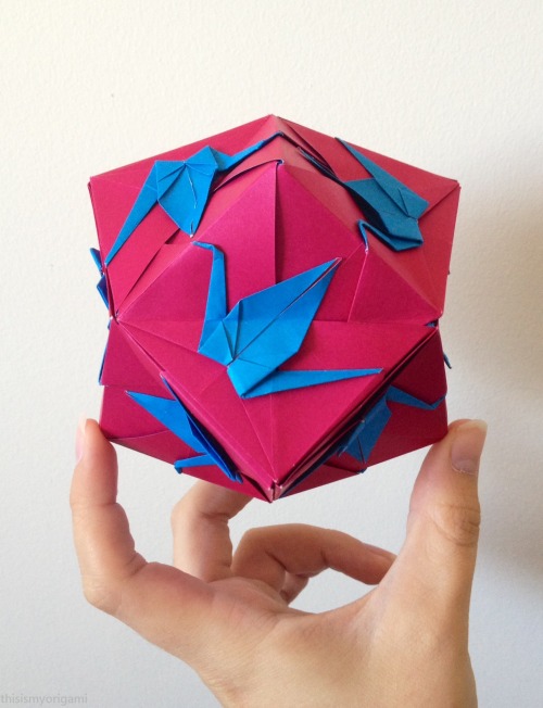
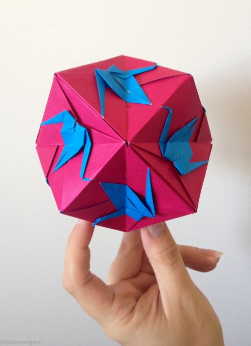
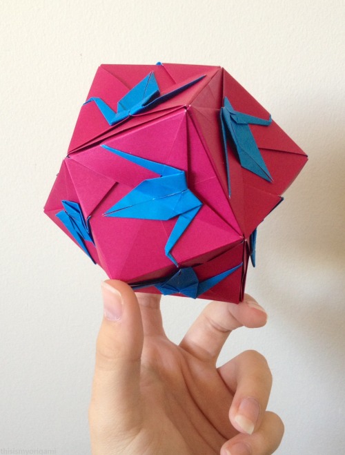
Crane Trisoctahedron designed by Fumiaki Shingu
Instructions
More Posts from Ritasakano and Others
The Great Conjunction of Jupiter and Saturn

Credits: NASA/Bill Ingalls
Have you noticed two bright objects in the sky getting closer together with each passing night? It’s Jupiter and Saturn doing a planetary dance that will result in the Great Conjunction on Dec. 21. On that day, Jupiter and Saturn will be right next to each other in the sky – the closest they have appeared in nearly 400 years!
Skywatching Tips from NASA

Credits: NASA/JPL-Caltech
For those who would like to see this phenomenon for themselves, here’s what to do:
Find a spot with an unobstructed view of the sky, such as a field or park. Jupiter and Saturn are bright, so they can be seen even from most cities.
An hour after sunset, look to the southwestern sky. Jupiter will look like a bright star and be easily visible. Saturn will be slightly fainter and will appear slightly above and to the left of Jupiter until December 21, when Jupiter will overtake it and they will reverse positions in the sky.
The planets can be seen with the unaided eye, but if you have binoculars or a small telescope, you may be able to see Jupiter’s four large moons orbiting the giant planet.
How to Photograph the Conjunction

Credits: NASA/Bill Dunford
Saturn and Jupiter are easy to see without special equipment, and can be photographed easily on DSLR cameras and many cell phone cameras. Here are a few tips and tricks:
These planets are visible in the early evening, and you’ll have about 1-2 hours from when they are visible, to when they set. A photo from the same location can look completely different just an hour later!
Using a tripod will help you hold your camera steady while taking longer exposures. If you don’t have a tripod, brace your camera against something – a tree, a fence, or a car can all serve as a tripod for a several-second exposure.
The crescent Moon will pass near Jupiter and Saturn a few days before the conjunction. Take advantage of it in your composition!
Get more tips HERE.
Still have questions about the Great Conjunction?
Our NASA expert answered questions from social media on an episode of NASA Science Live on Thursday, Dec. 17. Watch the recording HERE.
Make sure to follow us on Tumblr for your regular dose of space: http://nasa.tumblr.com.
https://www.instagram.com/rita.baanoai/
Babadores exclusiva feitos pela vovó Rita.


Os caquis maduros pintam o tempo cinza do inverno.
Caqui a fruta doce do inverno.


Ryohei Tanaka, Persimmons
Muitas vezes as imagens nos leva a mundos internos.

by Hisanori Manabe
Lindas imagens!
The Beautiful Things inside Your Head: Winners of the 10th Annual Art of Neuroscience Contest
Winner: Lidija Kononenko

Artist Kononenko described this interactive piece as “a microscope specimen, a map of symptoms, and an investigation of the unknown” in a statement accompanying it. Viewers can zoom in and explore the details of a microscope image of the peripheral nerve system, which is overlaid by textual facts and poetic phrases about sleep.Sleep is “a voluntary act of losing one’s own consciousness,” Konenenko explained in her statement. The poetic snippets resemble the fragmented thoughts humans have while falling asleep. And zooming in and out of the image represents the transition between wakefulness and sleep. Additionally, 31-3594 allows the viewer to act as a pathologist, achieving the goal of blending neuroscience and art. In assessing this unique piece, the jurors praised it for “the interactivity and playful combination of imagery of a human peripheral nerve with a text-based story that unfolds at various scales and highlights the role of the nervous system in the human condition.”
Honorable Mentions

Red Haze by Nicki Coveña
A tsunami of red dots dominates this image by neuroscientist Coveña. The bright red color comes from a fluorescent protein, which was used to visualize the workings of TBR1—a gene that synthesizes the protein that regulates the information transfer from DNA to messenger RNA in vertebrate embryo development. “The out-of-focus view makes one guess at what details are hidden below,” the jurors wrote.

Motor White Matter Networks of the Human Brain by Sanja Budisavljevic
In this piece, neuroscientist Budisavljevic superimposed color onto a 19th-century black-and-white drawing of a brain based on a postmortem dissection. Each color indicates a different “highway,” or white matter pathway connecting particular regions of gray matter and allowing information to be transferred. Red indicates the most prominent highway, which links the cortex and spinal cord. “This pathway carries the messages to and from the body and allows us to function in our sensory world,” Budisavljevic says. Green represents the connection that supports coordination, and blue shows the one that regulates movements.

Bdl by Paméla Simard (Alex Tran, photographs)
Artist Simard partnered with Hunter Shaw, a neuroscientist then at McGill University, to create a series of delicate wooden sculptures. “The various installations were created from fluorescent microscopy images representing the visual system of the fruit fly brain,” Simard wrote in her statement. The intricate details of the fruit fly visual system were made possible by first laminating the thin slices of different types of wood together, then hand cutting the result to mimic the microscope images.
More Art from 2020 Gallery

Whale Retina Rainbow by Elena Vecino Cordero and Luis López Vecino
In February 2019 the death of a whale in Sopelana Beach in Spain made the local news. The beach happened to be close to the University of the Basque Country, where biologist Vecino Cordero works. Seizing the opportunity, she and some volunteers extracted the eye of the whale and took it back to her ophthalmology research group for further study. The image was produced as a part of their research. The whale’s retina was imaged using scanning electron microscopy. And later López Vecino added the colors using Adobe Photoshop.

Sensing Spin by Dan Jagger
Physiologist Jagger used a high-resolution microscope to capture this image. It shows mechanosensory hair cells located in the inner ear that play a role in the sense of balance. A protein called actin is within bundles of stereocilia and is stained yellow. Actin helps the bundles to stand upright, so when the human head turns, they can detect the movement of the fluid they are immersed in. The hair-cell nuclei are stained with cyan.

The Protection of Nature Starts in Our Mind by Robert Luck
Luck is a neuroscientist at Heidelberg University in Germany who studies the development of the cerebellum, located where the spinal cord meets the brain. Alarmed by climate change and deforestation, he created a “mind forest” that resembles bird’s-eye-view photographs of real forests. The “trees” are 65 individually traced images of mice’s Purkinje neurons, which play important roles in controlling coordination and movements. “I chose the number 65 to represent the number of years needed for the rainforest to regrow and gain back at least 80% of its diversity,” Luck wrote in his statement. “[Sixty-five] years—a human lifetime!”

Memories and Patterns: Oligodendrocytes by Shanthi Chandrasekar
Oligodendrocytes are glial cells that support and insulate long neuronal axons. The cells’ lipid membrane wraps around the axons to strengthen the structure, as well as to help neurons to send signals quickly. “A single oligodendrocyte can connect with multiple axons,” artist Chandrasekar wrote in her statement. “In this [pen-and-ink] drawing, I have tried to bring out the connectedness of the oligodendrocytes and the axons.”

Shelter in Place by Geinene Carson
As its title suggests, this piece represents “the artist’s interpretation of the pandemic experience” while sheltering in place because of COVID-19, according to artist Carson’s statement. This acrylic-on-canvas piece is a part of a series entitled Neuron, which started as “visual prayers for our daughter with a rare genetic disorder,” Carson wrote on her Web site. While Shelter in Place implies physical restrictions, Carson, who is based in Atlanta, draws inspiration from the neural network, “because as important as our physical surroundings are to our state of living, our thought life holds the key to thriving within whatever the circumstances may be,” she wrote.

Bridges between Genesis and Neuroscience: Triplets by Rui Rodrigues
This image features three neurospheres—clusters of neural stem or progenitor cells—that are similar in size and shape. Because of their similarity, neurobiologist Rodrigues entitled the piece Triplets. The vibrant colors come from “antibodies coupled with fluorescent tags to label specific proteins,” he says.

The Transfer by Geinene Carson

Motor Neuron Architectural Digest by Stefanie Hauck - University of Bonn

Illuminating The Vascular Network - EPFL by Marwan Abdellah
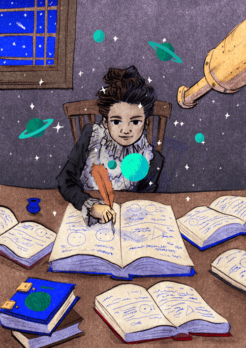
On this day but in 1750, Caroline Lucretia Herschel was born.
Caroline Herschel was the sister of the astronomer William Herschel. After learning astronomy alone and math with the help of her brother, she became his assistant. His most significant contribution to astronomy were the discoveries of various comets, especially comet 35P / Herschel-Rigollet.

-
 guipodongpisubs liked this · 1 year ago
 -
 t4tjunpei reblogged this · 3 years ago
 -
 trueger1 liked this · 4 years ago
 -
 kittybb liked this · 4 years ago
 -
 eugenia-virginia reblogged this · 5 years ago
 -
eugenia-virginia liked this · 5 years ago
-
 winkiieee23 liked this · 5 years ago
 -
 rramaret liked this · 5 years ago
 -
 the-heart-of-my-mystery reblogged this · 6 years ago
 -
 otto-von-stirlitz reblogged this · 6 years ago
 -
 addaenurse liked this · 6 years ago
 -
 nautilusopus liked this · 6 years ago
 -
 tocasia reblogged this · 6 years ago
 -
tocasia liked this · 6 years ago
-
 8sidedemi reblogged this · 7 years ago
 -
 buffoonfromtheblacklagoon reblogged this · 7 years ago
 -
 pepsirps reblogged this · 7 years ago
 -
 rosebouqu3t reblogged this · 7 years ago
 -
rosebouqu3t liked this · 7 years ago
-
 honestlyable-blog liked this · 7 years ago
 -
 giggling-breeze liked this · 8 years ago
 -
 myorizuru liked this · 8 years ago
 -
 ritasakano reblogged this · 8 years ago
 -
ritasakano liked this · 8 years ago
-
 giselujan-blog liked this · 8 years ago
 -
 camilaacriadora reblogged this · 8 years ago
 -
 nazneen-bandi liked this · 8 years ago
 -
 mendelmeups-blog liked this · 8 years ago
 -
 crepecrocodile liked this · 8 years ago
 -
 sharkbait-007-blog reblogged this · 8 years ago
 -
 pjpj-archived reblogged this · 8 years ago
 -
 juliathpiton-blog liked this · 9 years ago
 -
 yagyou liked this · 9 years ago
 -
 carotjuice liked this · 9 years ago
 -
 palejoke reblogged this · 9 years ago
 -
palejoke liked this · 9 years ago